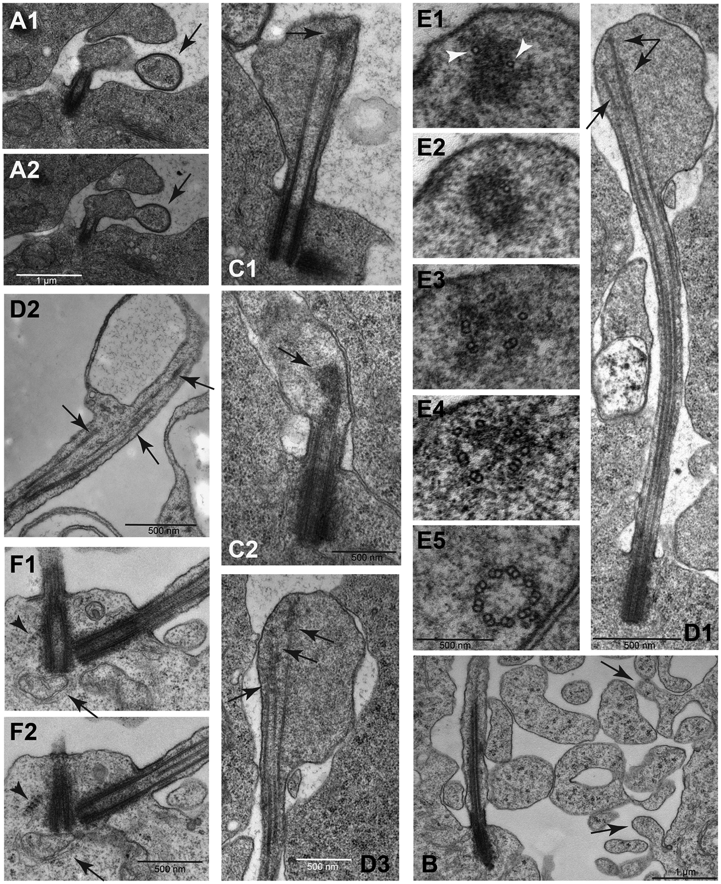
Fig. 4.

Unique aspects of cilium growth in Pieris spermatocytes. (A1–A2): Consecutive sections showing the contact of a large extracellular vesicle (arrow) with the growing cilium. (B): The extracellular space of the spermatocytes during the cilia growth phase is filled with free large vesicles containing ribosomes and distinct cytoplasmic blebs emerge from the cell membrane (arrows). (C1–C2): As prophase progresses the distal ends of the elongating cilia still maintain a distinct swelling where the microtubules end in a distal cluster of electrondense material (arrows). (D1–D3): The microtubules within the distal swellings have variable lengths (arrows); D3 is a close-up of a different section of the sample shown in D1. (E1–E5): Serial sections from the apex of a cilium to its base: single microtubules are associated with the dense material at the distal end of the cilia (arrowheads); moving apically toward the basal body, there are singlet microtubules, then scattered doublets, and at the base of the cilium doublets are arranged in a usual nine-fold symmetry. Central doublets and dynein arms are lacking in early stage cilia. (F1–F2): Striated structures resembling ciliary rootlets are found in close association with the mother centrioles in primary spermatocytes (arrowhead). Apparent mitochondria are seen at the base of the basal body (arrow).
