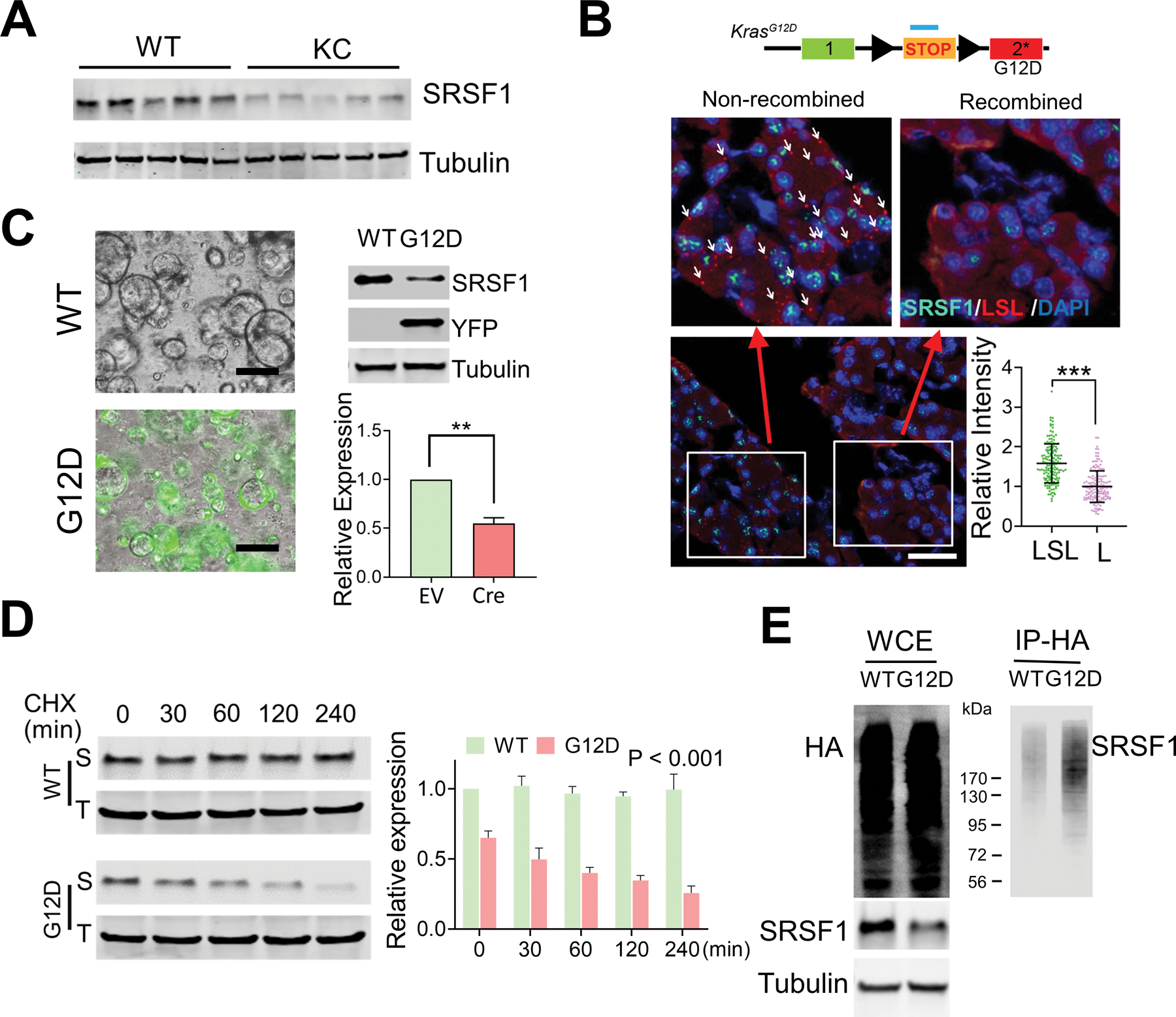
Figure 5.

Reduced expression of SRSF1 in morphologically normal KRASG12D-expressing pancreatic cells. A, Western blotting of SRSF1 protein in pancreatic protein lysates from WT and KC mice (n = 5 per group). B, Multiplexed RNA-FISH/IF staining of STOP-cassette and SRSF1 protein in pancreata isolated from KC mice. RNA FISH probes were designed to target the STOP cassette. Reflecting the mosaic expression of Cre recombinase in KC mice, pancreatic cells with the STOP cassette recombined and non-recombined can be distinguished by RNA-FISH probes. Upper left: non-recombined region, with white arrows indicating some of the positive FISH signals; upper right: recombined region, where most cells have undergone recombination and hence the FISH signal is lost; Bottom right, quantification of SRSF1 intensity in LSL-positive or -negative pancreas cells. Scale bars, 50 μm. Unpaired, two-tailed t test. ***p < 0.001. Error bars represent mean ± SD. C, YFP fluorescence (left), and western blotting and quantification of SRSF1 (right) of LSLKrasG12D/+; R26-LSl-YFP ductal organoids infected with adeno-empty (WT) or adeno-Cre (G12D) (n = 3 biological replicates). Unpaired, two-tailed t test. **p < 0.01. Error bars represent mean ± SD. D, Western blotting of SRSF1 (S) and Tubulin (T), and quantification in LSLKrasG12D/+; R26-LSl-YFP ductal organoids infected with adeno empty (WT) or adeno-Cre (G12D), respectively, followed by exposure to cycloheximide (10 mg/ml) for the indicated times. Linear mixed effects model with the experimental group, time and group by time interaction as fixed effects, and sample-specific random intercept was used to estimate the relative expression. E, In vivo ubiquitination assay of HA-ubiquitin stable-expressing LSL-KRASG12D/+; R26LSL-YFP organoids infected with adeno empty (WT) and adeno-Cre (G12D), respectively. Following immunoprecipitation of cell lysates, immunoprecipitates (IP) and whole-cell extracts (WCE) were analyzed by immunoblotting with the indicated antibodies.
