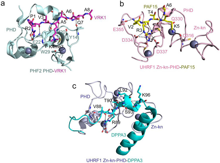
Figure 1: H3 mimetics occupy the canonical histone binding pocket.
(a) A ribbon diagram of the crystal structure of the PHF2 PHD finger in complex with the histone-mimicking peptide derived from the VRK1 protein (PDB ID 7M10). The VRK1 peptide is shown as magenta sticks, and the zinc ions are grey spheres. Intermolecular hydrogen bonds are indicated by black dash lines. (b) The crystal structure of the zinc-knuckle-PHD finger of UHRF1 in complex with PAF15 peptide (PDB ID 6IIW). The PAF15 peptide is shown as yellow sticks, and intermolecular hydrogen bonds are indicated by black dash lines. (c) The solution NMR structure of the zinc-knuckle-PHD finger of UHRF1 linked with DPPA3 (Stella) (PDB ID 7XGA). Hydrogen bonds between the UHRF1 (light blue) and DPPA3 (cyan) residues are indicated by black dash lines.

