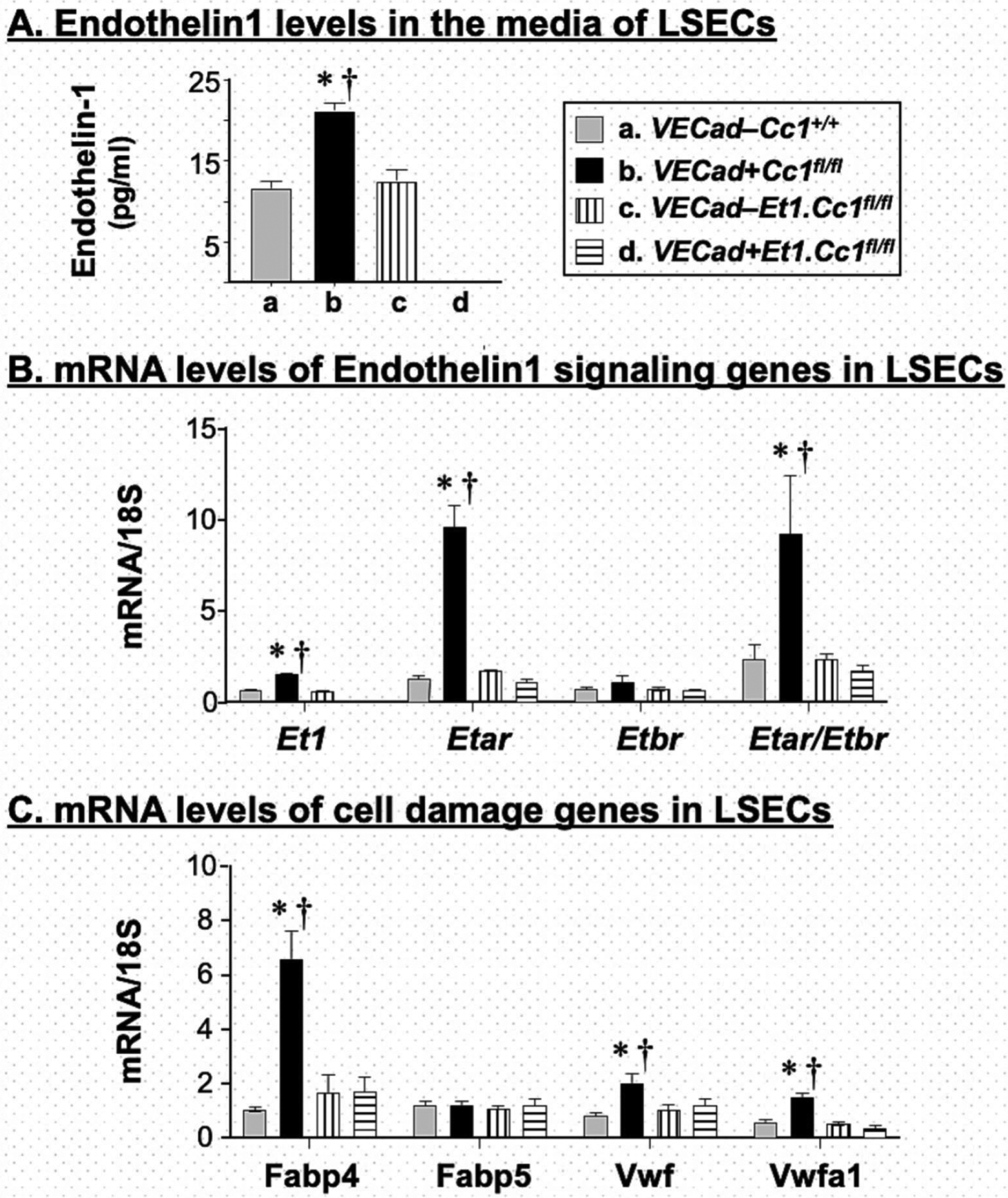
Fig. 7. Analysis of liver sinusoidal endothelial cells (LSECs).

LSECs were isolated from mice at 2–3 months of mice, allowed to grow for 3 days in 3 separate wells of 24-well plates before (A) the media was collected to assess ET1 levels and cells were lysed for qRT-PCR analysis of mRNA levels of genes involved in (B) ET1 signaling and (C) cell damage. Measurements were done in duplicate. Values were expressed as mean ± SEM. *P<0.05 vs control/each genotype and †P<0.05 VECad+Cc1fl/fl.ET1fl/fl vs VECad+Cc1fl/fl.
