Figure 6.

SIRT7 promotes NSC maintenance by suppressing the mitochondrial protein folding stress.
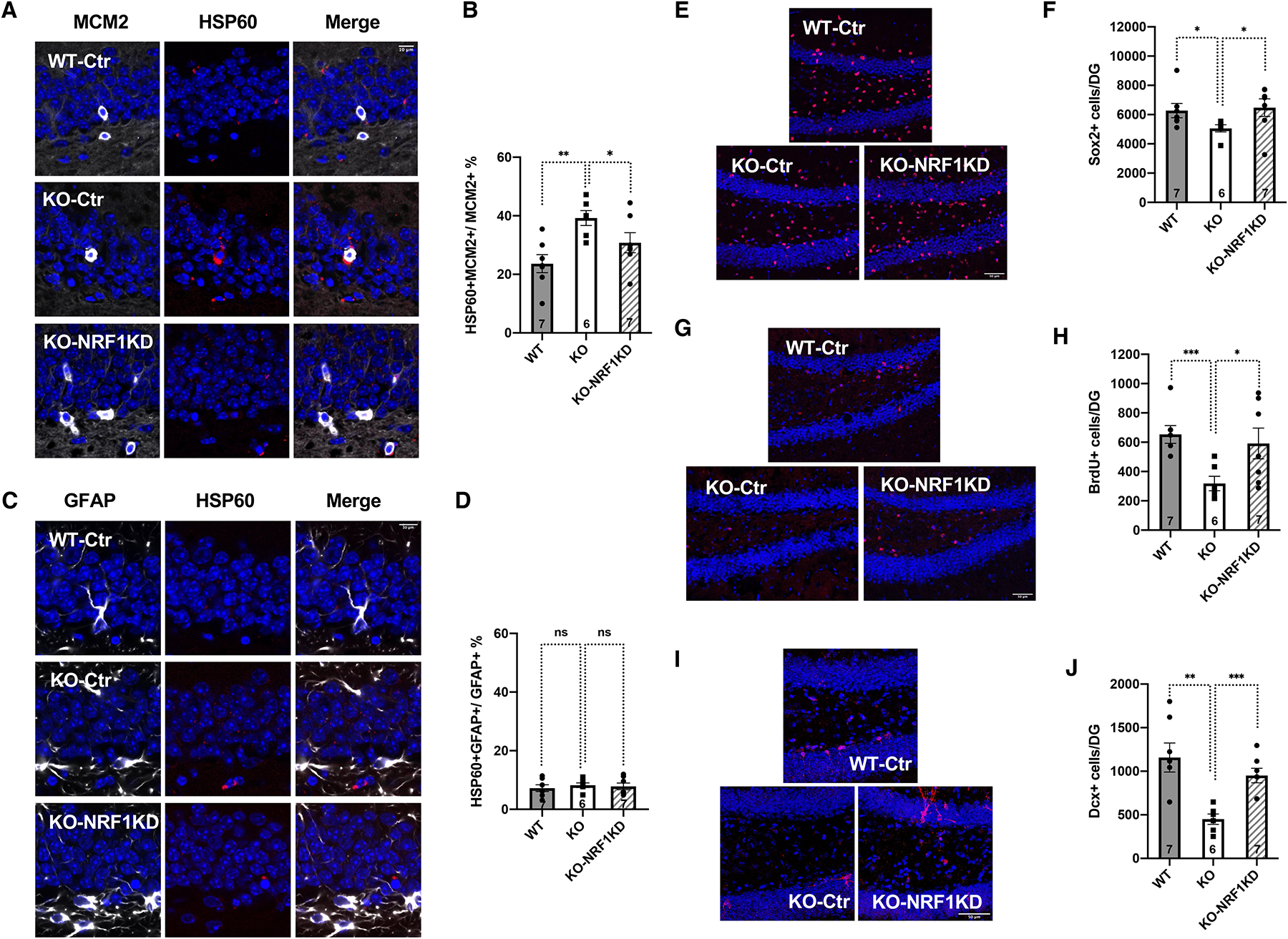
Comparison of 6–7-month-old WT and SIRT7−/− mice that were given stereotaxic injections of control lentivirus or lentivirus knocking down NRF1 into the dentate gyrus.
A-D, Immunohistochemistry staining and quantification of MCM2 and HSP60 (A, B) and GFAP and HSP60 (C, D) in the dentate gyrus. Blue: DAPI. Red: HSP60. White: MCM2 or GFAP. Scale bar: 10 μm.
E, F, Immunohistochemistry staining (E) and quantification (F) of Sox2-positive cells in the dentate gyrus. Blue: DAPI. Red: Sox2. Scale bar: 50 μm.
G, H, Immunohistochemistry staining (G) and quantification (H) of long-term BrdU-retaining cells in the dentate gyrus. Blue: DAPI. Red: BrdU. Scale bar: 50 μm.
I, J, Immunohistochemistry staining (I) and quantification (J) of newly differentiated Dcx-positive neurons in the dentate gyrus. Blue: DAPI. Red: Dcx. Scale bar: 50 μm.
Error bars represent SE. *: p<0.05. **: p < 0.01. ***: p<0.001. ns: p>0.05. Student’s t test. See also Figure S13, S14, S15, Video S3.
