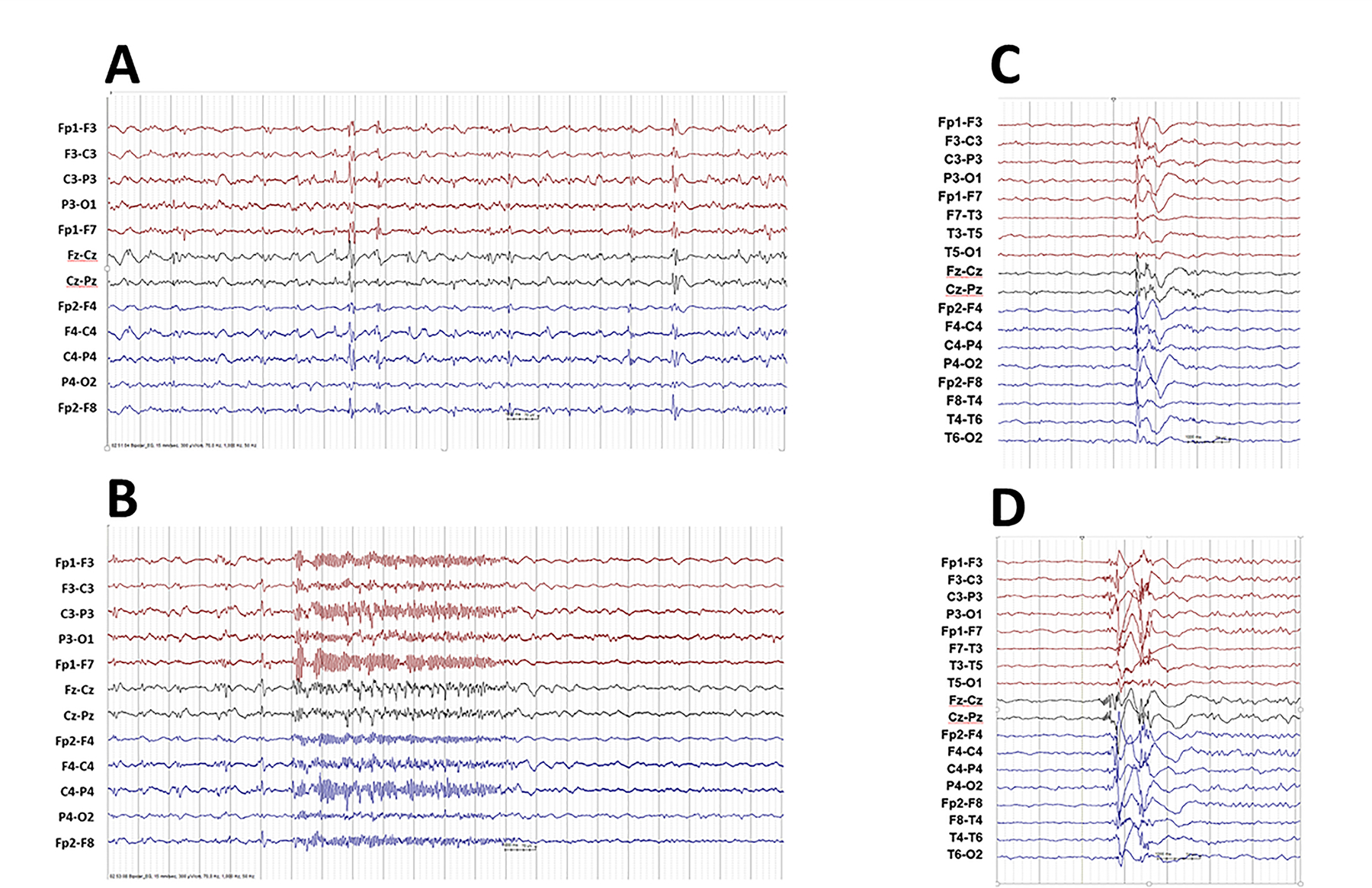
Figure 1A-B; C-D.

A-B. Individual 15. Interictal EEG showing runs of irregular delta activities with diffuse spike-slow wave complexes and background slowing. Ictal EEG showing diffuse recruiting rhythmic fast discharge associated with a tonic seizure.
C-D. Individual 10. EEG recorded during sleep showed generalized epileptiform discharge of polyspike-and-slow-wave (C) originating from right frontal /centro-frontal followed by alpha-activity with discrete right-sided prevalence, and (D) originating from the vertex followed by alpha-activity with discrete right-sided prevalence.
