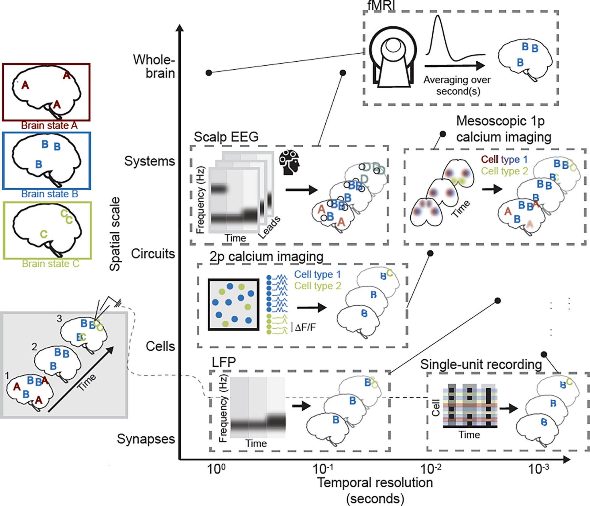
Figure 1. Systems and cognitive neuroscience techniques each offer partial, but complementary, insight into brain states.

In this example, three schematic brain states, A, B, and C, are depicted (upper left). Each brain state is defined by a stereotyped pattern of whole-brain activity, and brain states can occur in isolation (timepoint 2; bottom left) or in combination (timepoints 1 and 3). Commonly used techniques are schematized and placed (see corresponding point) according to their approximate, relative temporal resolution and spatial scale (i.e., field of view size), as typically implemented. Single-unit recording: Schematized raster plot represents cell spiking at three time points. Limited field of view is represented by focal patterns, and cell response heterogeneity that may obscure patterns is represented by incomplete “B” and “C”. The identity of the cells is typically unknown. Two-photon (2p) calcium imaging suffers from many of the same limitations (represented by focal, partial brain state patterns and heterogeneous calcium signal at the individual cell level), but can offer insight into cell-type specificity of brain state expression (represented by colored cells, each of which preferentially responds during the brain state of that color). Local field potentials (LFPs) capture regional fluctuations in activity with high temporal resolution, and can be used, for instance, to assess changes in frequency of extracellular oscillations across brain states. Mesoscopic single-photon (1p) calcium imaging has slightly lower temporal resolution, but captures cell type-specific patterns of brain activity across the cortical mantle, yielding more complete representations of brain states (though with limited resolution of activity in deep structures, represented by blurred pattern elements). Scalp electroencephalography (EEG), like LFPs, captures brain state-associated differences in power across frequency bands, but suffers from imprecise source localization (represented by enlarged, blurred, and conflated pattern elements – i.e., pattern D that merges brain states B and C). Functional magnetic resonance imaging (fMRI), like EEG, offers whole-brain coverage in humans, but has low temporal resolution, such that brain state patterns will be effectively averaged over seconds. In this example, the only resolved brain state will thus be brain state B, which persists across all time points.
