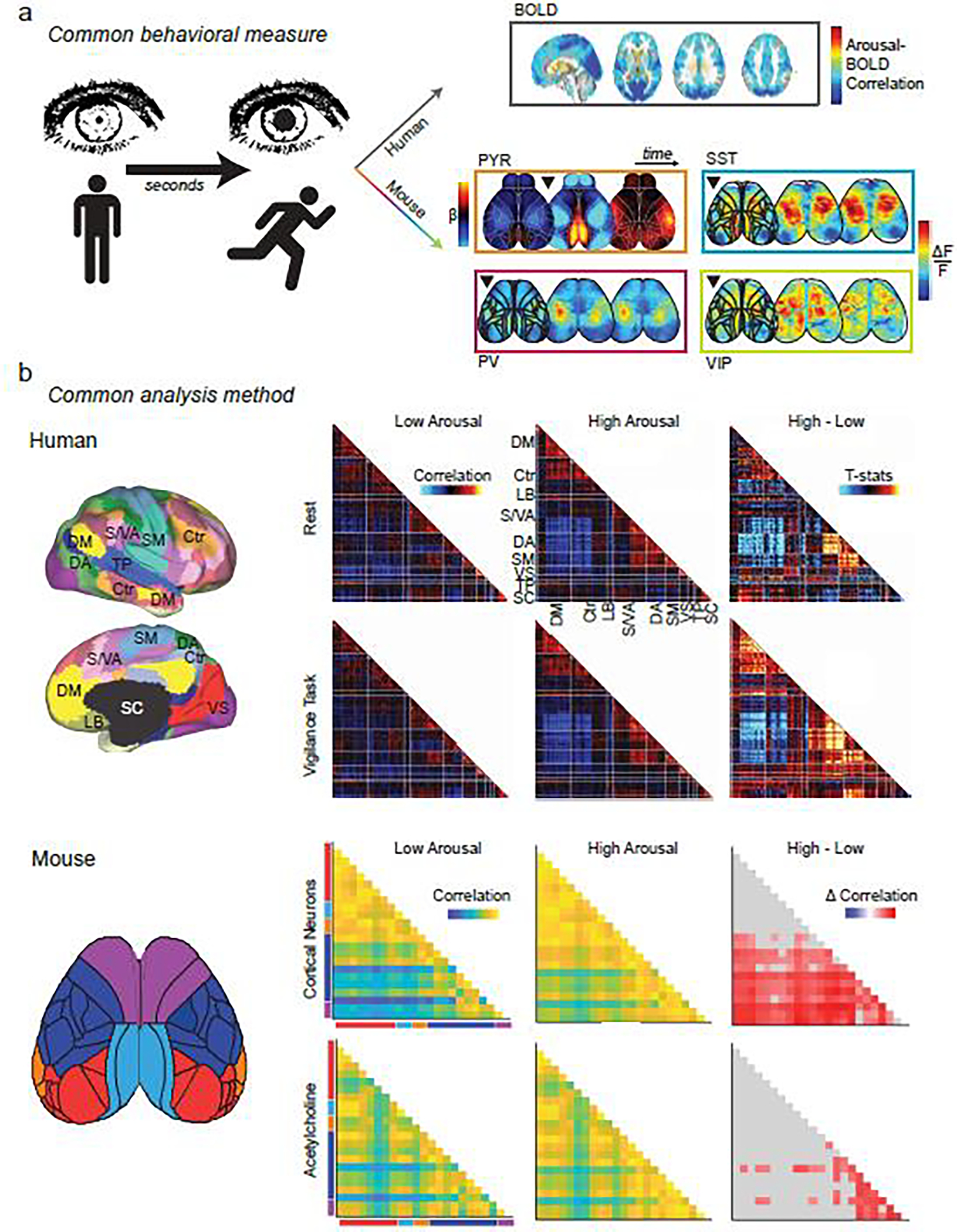
Figure 4. Using brain state to unify the study of neural dynamics.

Common behavioral measures link the study of brain states across species: a, Changes in arousal measures (i.e. pupil size, movement) occur on the order of seconds. Top right: the slow dynamics of BOLD fluctuations on this time scale yield persistent (but whole-brain) patterns related to changes in arousal. Adapted from [159]. Bottom right: faster dynamics (each image represents changes over ~300 msec) can be captured with cell-type specificity using mesoscopic calcium imaging in mice. Adapted from [37] (PYR) and [66] (PV, SST, VIP). Black triangle represents onset of increased arousal. By matching behavioral states across species, more invasive, higher resolution techniques in animal models can be leveraged to reveal mechanistic insights into the complex behavioral and cognitive processes that can uniquely be studied in humans. Shared analytic methods also provide a tool to bridge disciplines: b, Whole-brain activity patterns can be divided into parcels within functionally defined regions (left) and pairwise correlations of parcels’ time courses yield functional connectivity (FC) matrices, whether from fMRI data in humans (left top) or from mesoscopic functional imaging data in mice (left bottom). FC can be calculated across physiological states (low versus high arousal) from data acquired during a given cognitive state (e.g. rest versus task) or from measures of neuronal activity or acetylcholine release. These FC matrices can be used to reveal common features of low- and high-arousal brain state patterns and yield mechanistic insight into how these states may be generated. For example, tasks amplify arousal-related FC increases in somato-motor networks in humans (right top and upper middle matrices). Adapted from [160]. Increases in arousal also cause correlated acetylcholine release and synchronize neuronal activity in analogous networks in mice (right lower middle and bottom matrices). Adapted from [27]. Human network labels: DM, default mode; Ctr: control; LB: limbic; S/VA: salience/ventral attention; DA: dorsal attention; SM: somato-motor; VS: visual; TP: temporo-parietal; SC: subcortical. Mouse network labels: red: visual; light blue: retrosplenial; orange: auditory; dark blue: somatosensory; purple: motor.
