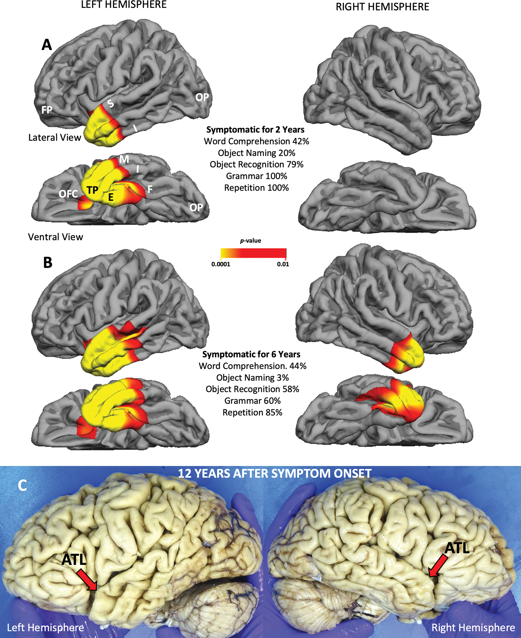
Figure 1. Selectivity of TDP-C for ATL Shown by Free Surfer and at Autopsy.

All images are from a semantic PPA patient (right-handed woman) with symptom onset at the age of 59 and TDP-C at autopsy. Significant atrophy on the cortical surface in A and B is shown by the colored areas, at the false discovery rate of 0.05 using the Free Surfer tool kit. A. Atrophy is confined to left ATL and is sufficient to cause severe and isolated impairment of word comprehension and object naming. B. Four years later all significant atrophy is still confined to ATL but has also emerged on the right. Object naming further decreased and non-verbal object recognition started to fail, likely due to bilaterality of ATL atrophy. C. Autopsy specimen of the patient 12 years after symptom onset. The macroscopic neurodegeneration is still strongly selective for ATL. Abbreviations: ATL-anterior temporal lobe; E- entorhinal/perirhinal cortex; F-fusiform gyrus; FP- frontal pole; I-inferior temporal gyrus; M- middle temporal gyrus; OFC- orbitofrontal cortex; OP-occipital pole; TP- temporal pole.
