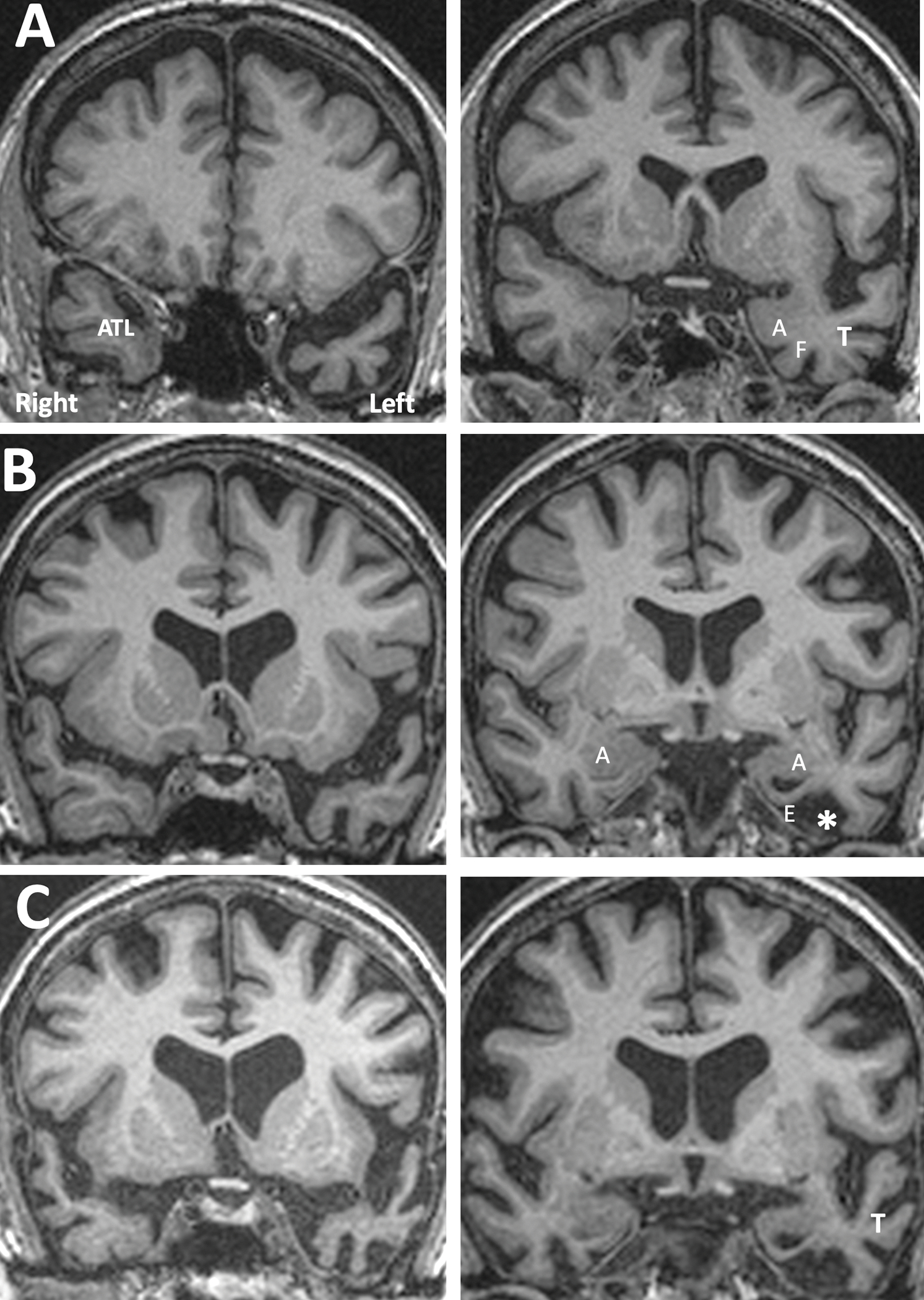
Figure 3. MRI Appearance of TDP-C Progression Illustrated by Individual Cases.

A. Distribution of atrophy at an early symptomatic stage. From a TDP-C semantic PPA patient (right-handed man) with onset at the age of 59. Significant atrophy is confined to the part of the left anterior temporal lobe (ATL) located ahead of the limen insulae. The amygdala, rhinal cortices, the fusiform gyrus, and lateral parts of temporal cortex are relatively preserved. B. Intermediate stage of atrophy from a right-handed woman with symptom onset at 59 (same patient shown in Figure 1). Atrophy of the left ATL has become more extensive. The amygdala has mild atrophy, rhinal cortices are severely thinned, the anteriormost part of the fusiform gyrus has been replaced by cerebrospinal fluid (*), and the temporal horn shows ex vacuo enlargement. C. A more advanced stage of atrophy. There is further neurodegeneration in left ATL, amygdala, rhinal cortices and the fusiform gyrus. Atrophy has now spread to the lateral temporal cortices (T). Abbreviations: A- amygdala; ATL- anterior temporal lobe; E- entorhinal/perirhinal areas; F- fusiform gyrus; T- lateral temporal cortices.
