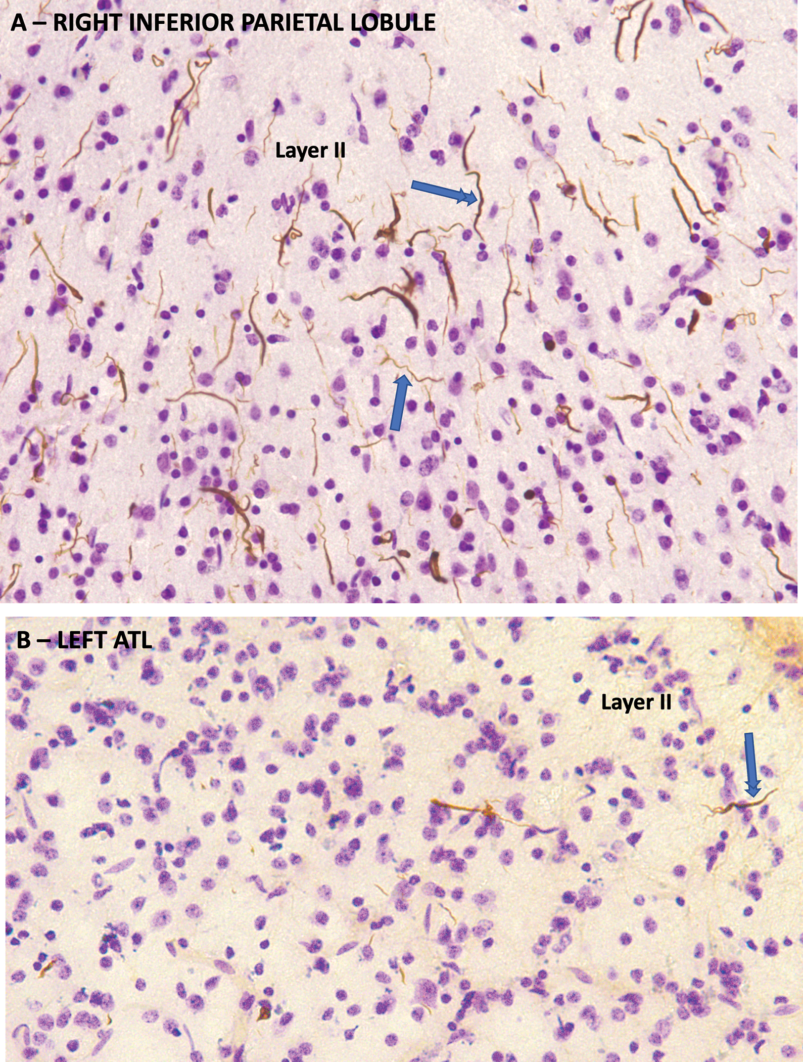
Figure 4. TDP-C Neuropathology.

TDP-43 immunohistochemistry counterstained with cresyl violet from the autopsy of a semantic PPA case with symptom onset at the age of 63 and death 10 years later. A. From the right inferior parietal lobule which was relatively spared. Neuronal architecture is mostly preserved. There is a high density of thick, long, dystrophic neurites pathognomonic of TDP-C, many of which have a rectilinear orientation perpendicular to the cortical surface in an orientation consistent with apical dendrites of pyramidal neurons (double arrowhead). Others are wispy and undulating, reminiscent of interneuron dendrites (single arrowhead). B. Same method showing the left ATL, which had advanced neurodegeneration. There is severe gliosis and loss of neurons. The density of dystrophic neurites is much lower than in the inferior parietal lobule.
