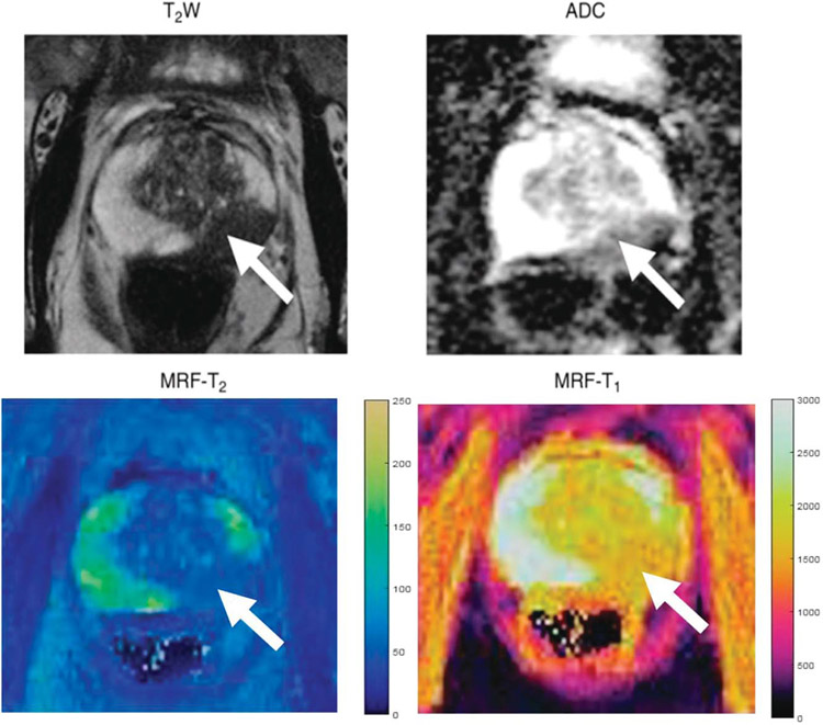
FIGURE 2.
Images in a 72-year-old man referred for elevated prostate-specific antigen level of 9.87 ng/mL with minimal urinary symptoms who underwent limited MR imaging and targeted biopsy of lesion in left mid prostate. Prostate adenocarcinoma with Gleason score 4 + 3 = 7 was diagnosed at cognitively targeted biopsy. T2-weighted image, ADC map, MRF T2 map, and MRF T1 map show corresponding hypointense lesion in mid prostate (arrow) and NPZ in right hemiprostate. Figure adapted with permission from Yu et al.94

