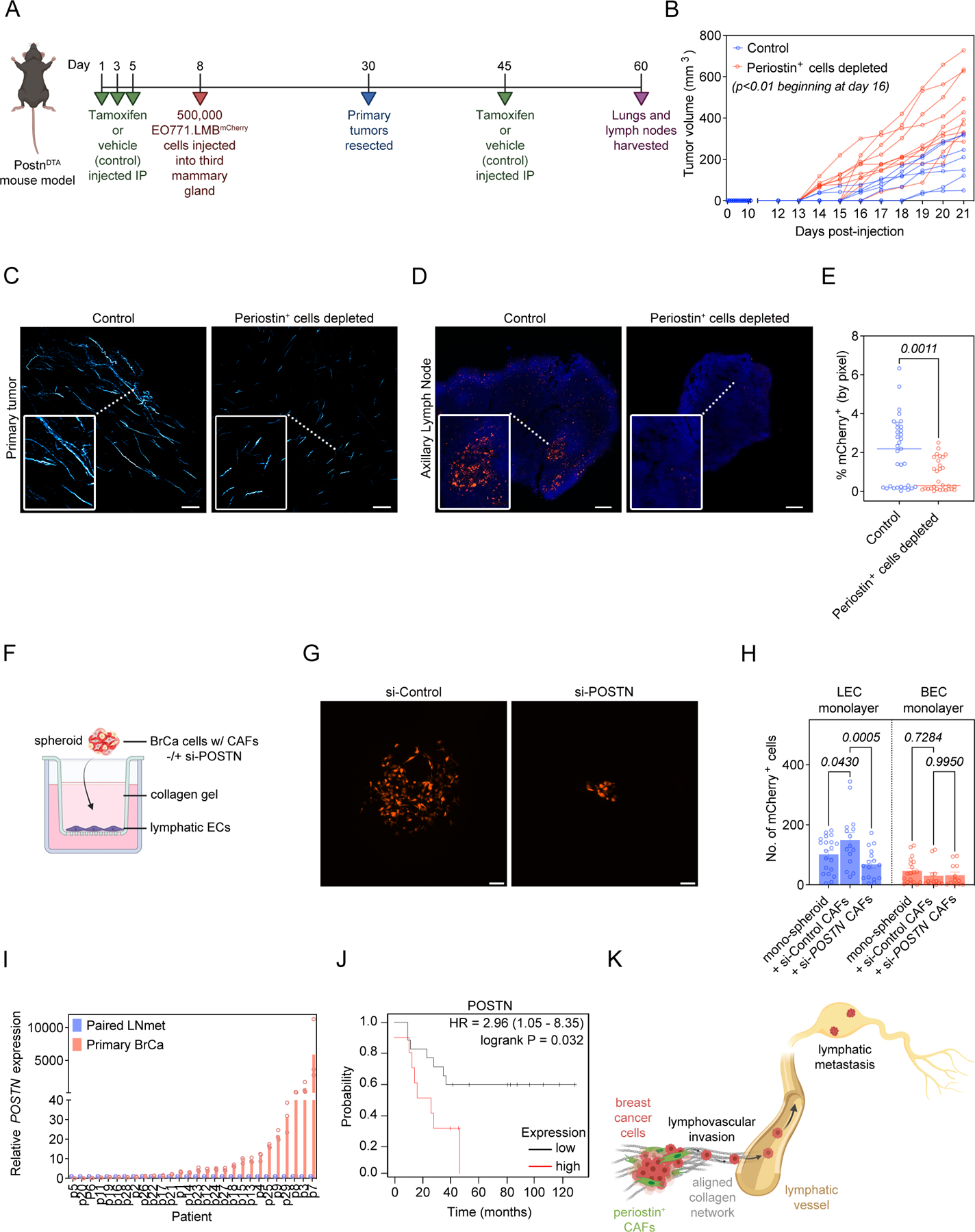
Figure 7. Periostin-expressing CAFs promote lymphatic metastasis by remodeling the extracellular matrix and directing lymphovascular invasion along organized collagen fibers.

(A) Study design in PostnDTA mice. (B) Volume measurements of EO771.LMB tumors in control versus periostin+ cell-depleted mice. Each line represents an individual mouse (n = 8–10 mice per group). Statistics shown for multiple Mann-Whitney tests. (C) SHG images of intratumoral collagen fibers (pseudo-colored in cyan) in control versus periostin+ cell-depleted mice. Scale bars: 150 μm, insets: 3x zoom. (D) Tissue tilescans of axillary lymph nodes from control versus periostin+ cell-depleted mice bearing EO771.LMB mammary tumors. Tumor cells labelled with mCherry and nuclei counterstained with DAPI. Scale bars: 200 μm, insets: 3x zoom. (E) Percentage of tissue area positive for mCherry in sections of axillary lymph nodes from control versus periostin+ cell-depleted mice bearing highly metastatic EO771.LMB mammary tumors. Each data point represents an individual histological section (n = 8–10 mice per group). Statistics shown for Mann-Whitney test. (F) Experimental setup for in vitro lymphovascular invasion assay using MDA-MB-231mCherry human breast cancer cells, primary human breast CAFs, and primary human lymphatic endothelial cells. (G) Representative images of MDA-MB-231mCherry tumor cells that have invaded across the lymphatic endothelial cell barrier in the in vitro lymphovascular invasion assay. Scale bar: 100 μm. (H) Quantification of MDA-MB-231mCherry tumor cells that have invaded across the lymphatic endothelial cell (LEC) barrier or blood endothelial cell (BEC) barrier in the lymphovascular invasion assay. Spheroids consisted of either MDA-MB-231mCherry tumor cells alone (mono-spheroids) or co-cultures of MDA-MB-231mCherry tumor cells with primary human breast CAFs treated with either non-targeting control siRNA (si-Control) or periostin-targeting siRNA (si-POSTN). Each data point represents an individual spheroid. Experiment performed in triplicate. Statistics shown for 2-way ANOVA. (I) RT-qPCR analysis of periostin expression in paired primary breast cancer specimens (Primary BrCa) and lymph node metastases (Paired LNmet) from human breast cancer patients (n = 28 patients). qPCR performed in triplicate. (J) Kaplan-Meier plot of overall survival in lymph node positive breast cancer patients stratified by periostin protein expression (n = 27 patients). (K) Summary model. (Schematics created with BioRender.com).
