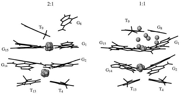
Figure 6.
The structure of the 2:1 complex is shown on the left. The structure of the DNA is that obtained at 100 ps in the trajectory. Only some of the bases of the DNA are shown for clarity. The spheres indicate the positions of the potassiums that were found for each of the 10 structures extracted from the trajectory from 80 to 100 ps. The spheres have one-third, 0.65 Å, the van der Waals radius of potassium, 1.96 Å. The analogous structures are shown on the right for the 1:1 case.

