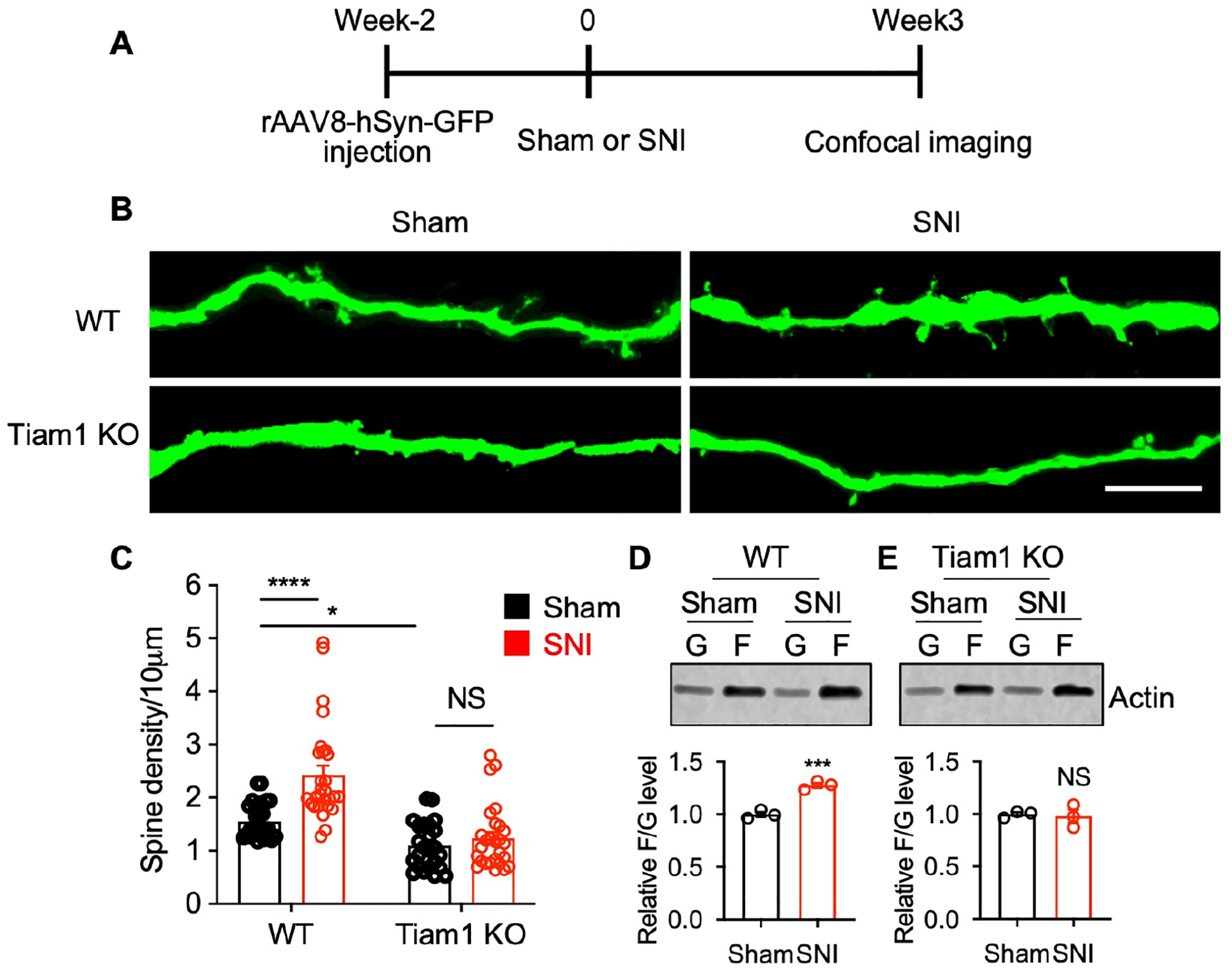
Figure 4. Tiam1 mediates synaptic structural plasticity in neuropathic pain.

(A) Experimental paradigm for dendritic spine analysis.
(B and C) Representative confocal images and quantification of dendritic spine density showing the increase in dendritic spines in dorsal horn spinal neurons following SNI in WT mice, but not global Tiam1 KO mice (scale bar, 10 μm). Two-way ANOVA analysis followed by Dunnett’s post-hoc test (n = 24–28 neurons from 3 mice/group. P < 0.0001, F1,103 = 47.77).
(D and E) Tiam1 deletion attenuates nerve injury-stimulated actin polymerization in the spinal dorsal horn. Western blots and quantification revealed that SNI increased the F- to G-actin ratio in the spinal dorsal horn of WT mice (D), but not global Tiam1 KO mice (E). Mann-Whitney U-test (n = 3).
Data are presented as means ± s.e.m. *P < 0.05, ***P < 0.001, ****P < 0.0001, NS, no significant difference.
See also Figure S7.
