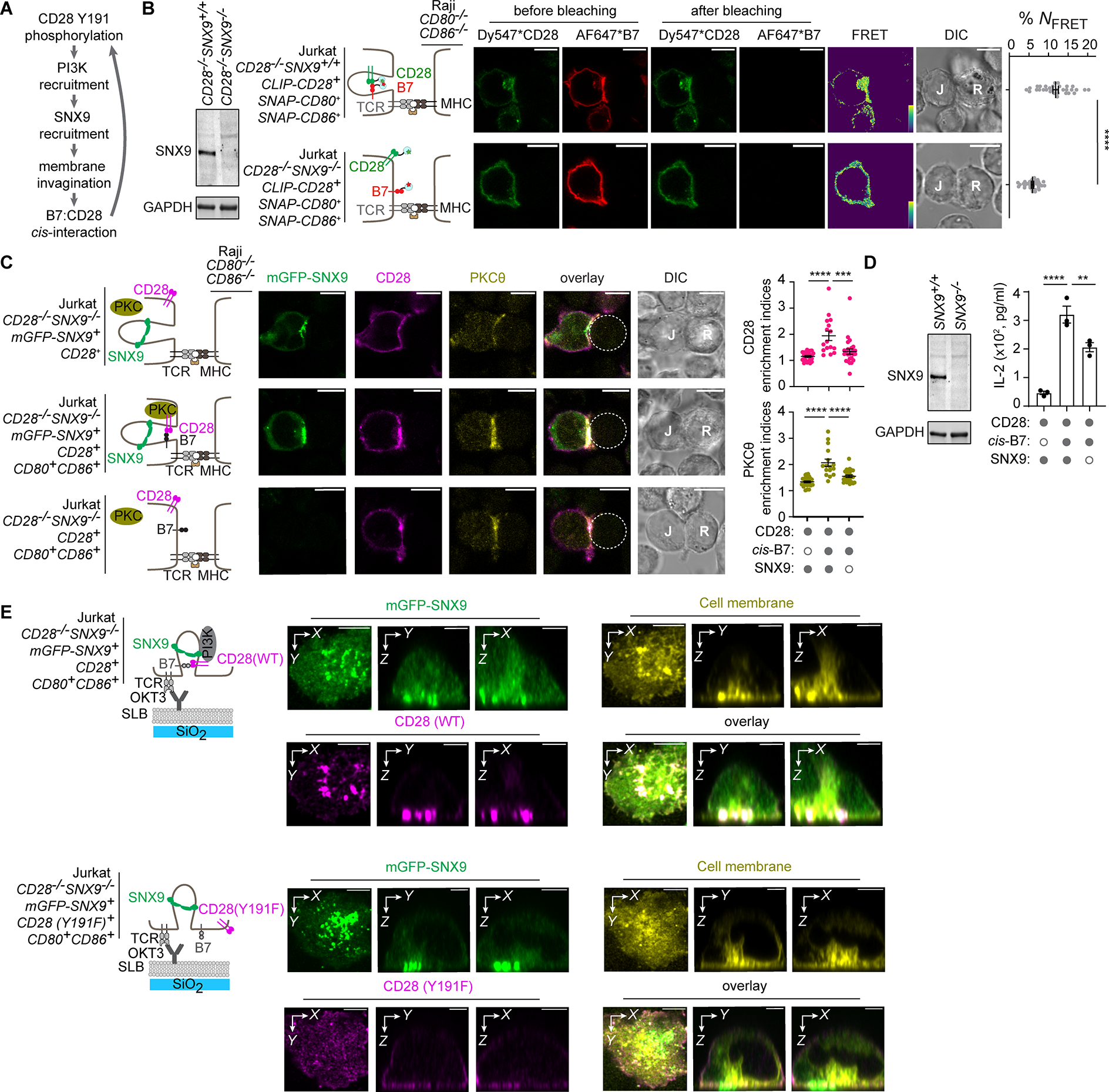
Figure 6. SNX9-induced membrane invagination at the T:APC interface promotes cis-B7:CD28 interactions and T cell activation.

(A) Model by which CD28:PI3K interaction promotes cis-B7:CD28 interactions via SNX9-driven membrane invagination.
(B) A FRET assay showing effects of SNX9 deficiency on cis-B7:CD28 interactions. Leftmost western blot shows SNX9 and GAPDH expression in indicated Jurkat cells. Immediate right are cartoons showing indicated Jurkat cells conjugated with CD80−/−CD86−/− Raji cell. Further right are confocal images of Dy547 and AF647 channels before and after AF647 photobleaching, FRET efficiency image, and DIC image. Bar = 10 μm. Dot plot: cis-B7:CD28 NFRET from n > 30 cells in three independent experiments. Mean ± SEM.
(C) Effects of SNX9 deficiency on cis-B7-induced synaptic enrichment of CD28 and PKCθ. Leftmost cartoons depict a CD80−/−CD86−/− Raji cell contacting Jurkat cell with indicated genotype. Further right are confocal images of immunostained CD28 and PKCθ acquired 30 min after Jurkat:Raji cell contact, with Raji cell denoted by a dash circle. Dot plots: synaptic enrichment indices of CD28 (magenta) and PKCθ (yellow), n > 16 conjugates in three independent experiments. Bar = 10 μm.
(D) Effects of SNX9 deficiency on cis-B7 induced IL-2 secretion. Western blot shows SNX9 and GAPDH expression in indicated Jurkat cells. Bar graph on the right summarizes IL-2 secretion from WT or SNX9−/− Jurkat cell with or without B7 transduced upon coculture with excess SEE-loaded CD80−/−CD86−/− Raji cell in three independent experiments.
(E) 3D reconstructed Z-stack confocal images of SNX9, T cell membranes, and CD28 in the presence of cis-B7 upon TCR stimulation. Leftmost cartoons depict Jurkat cell with indicated genotype triggered by an anti-CD3ε (OKT3) containing SLB. On the right are 3D reconstruction images of mGFP-SNX9, R18-stained Jurkat cell membranes, and immunostained CD28(WT) or CD28(Y191F) acquired 30 min after Jurkat:SLB contact. Bar = 5 μm.
Unpaired two-tailed Student’s t test for (B), One-way ANOVA for (C, D). **p < 0.01, ***p < 0.001, ****p < 0.0001.
