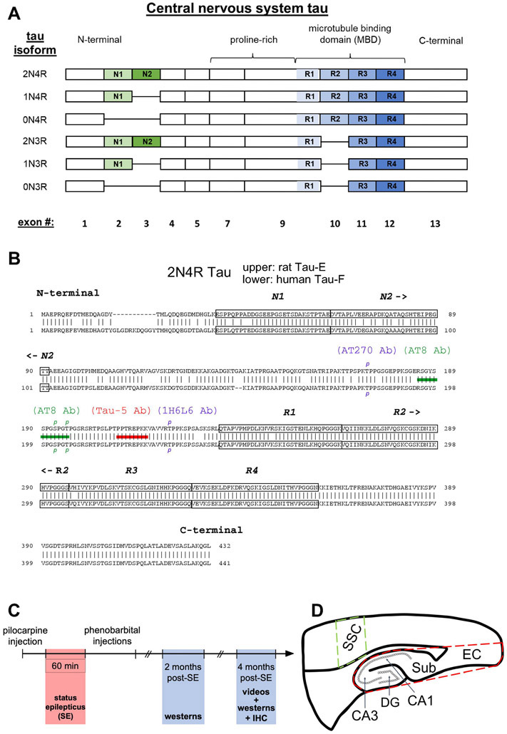
Figure 1.
Tau isoform composition. (A) Schematics of the six tau isoforms formed by alternative splicing of N-terminal exons (N1, N2, green-shaded) and of repeat domains (R1-R4, blue-shaded) within the microtubule binding region (MBD). The presence of none, one, or two N-terminal exons determines 0N, 1N, 2N tau isoform specificity, correspondingly; while the absence or presence of the R2 domain within the MBD generates 3R or 4R tau isoforms, respectively. Labeling at bottom is the exon number of the human microtubule associated protein tau gene, MAPT, that encodes for the corresponding domain above. Exons 6 and 8 are not expressed in the central nervous system. (B) Comparison of 2N4R tau amino acid sequences between rat (upper) and human (lower) showing 89% homology between the two species. Tau naming for each species found in Uniprot.org. The two N-terminal exons and four repeat domains are boxed within the sequences. Shown are epitopes for anti-tau antibodies (Abs) Tau-5 Ab (red bar) and AT8 Ab (green bar). To be recognized by the AT8 Ab, both S193 and T196 rat tau residues (S202 and T205 in humans) must be phosphorylated, denoted by green p’s. Additionally, two other phosphorylated tau residues, T172 rat/T181 human recognized by the AT270 Ab and T222 rat/T231 human recognized by the 1H6L6 Ab, are labelled as blue p’s. (C) Timeline of experiments involving the pilocarpine-induced rat model of chronic epilepsy. (D) Schematic of brain regions studied. Whole hippocampal formation encompassing CA1 and CA3 hippocampus, dentate gyrus (DG), subiculum (Sub), and entorhinal cortex (EC) is enclosed within the dashed red border. A region within the somatosensory cortex (SSC, dashed green boundary) served as control tissue outside the putative seizure onset zone within the hippocampus.

