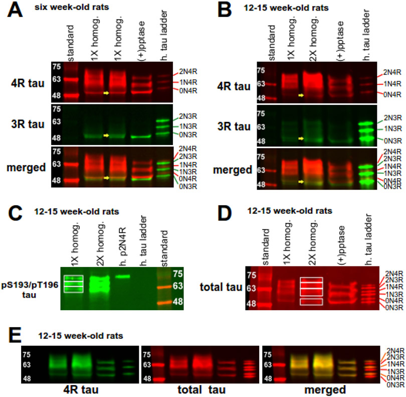
Figure 2.
Expression of tau isoforms in hippocampal tissue. A ladder containing six human tau isoforms (h. tau ladder) allows for size comparison and serves as a negative control for phosphorylation. (A) 4R and 3R tau expression in raw tissue homogenate either without (1X homog.) or with phosphatase treatment [(+) pptase] from six week-old naïve rats. In the raw homogenate lanes of the top panel displaying 4R tau isoforms, tau proteins migrate between 50 to 72 kDa. Consolidation of these isoforms into three protein bands in phosphatase-treated homogenates indicate 4R tau isoforms are highly phosphorylated. In the raw homogenate lanes displaying 3R tau (middle panel), a doublet around 50 kDa converged into the lower band after phosphatase treatment. The bottom panel is a merged image showing four main tau isoforms. The yellow arrows in the three panels demonstrate that the upper doublet band in the merged panel consists of 0N3R tau and 0N4R tau. (B) 4R and 3R tau expression in 12-15 week-old naïve rats. Tau expression is similar as above, except for notably decreased expression of both 0N3R and 0N4R isoforms in the older rats. (C) AT8 Ab staining of dually phosphorylated S193/T196 tau in brain homogenates from 12-15 week old rats. Three tau bands are observed. Phosphorylated 2N4R human tau (h. p2N4R) served as a positive control for detection of dually phosphorylated S193/T196 tau. (D) Total tau staining of rat brain homogenates with the Tau-5 Ab. Four bands of staining are observed in raw homogenates. Phosphatase treatment consolidates staining to three bands. (E) Complete overlapping (merged) of bands composed of 4R tau (4R tau, green) and total tau (total tau, red) demonstrates that total tau staining reflects 4R tau expression in brain homogenates of 12-15 week-old rats.

