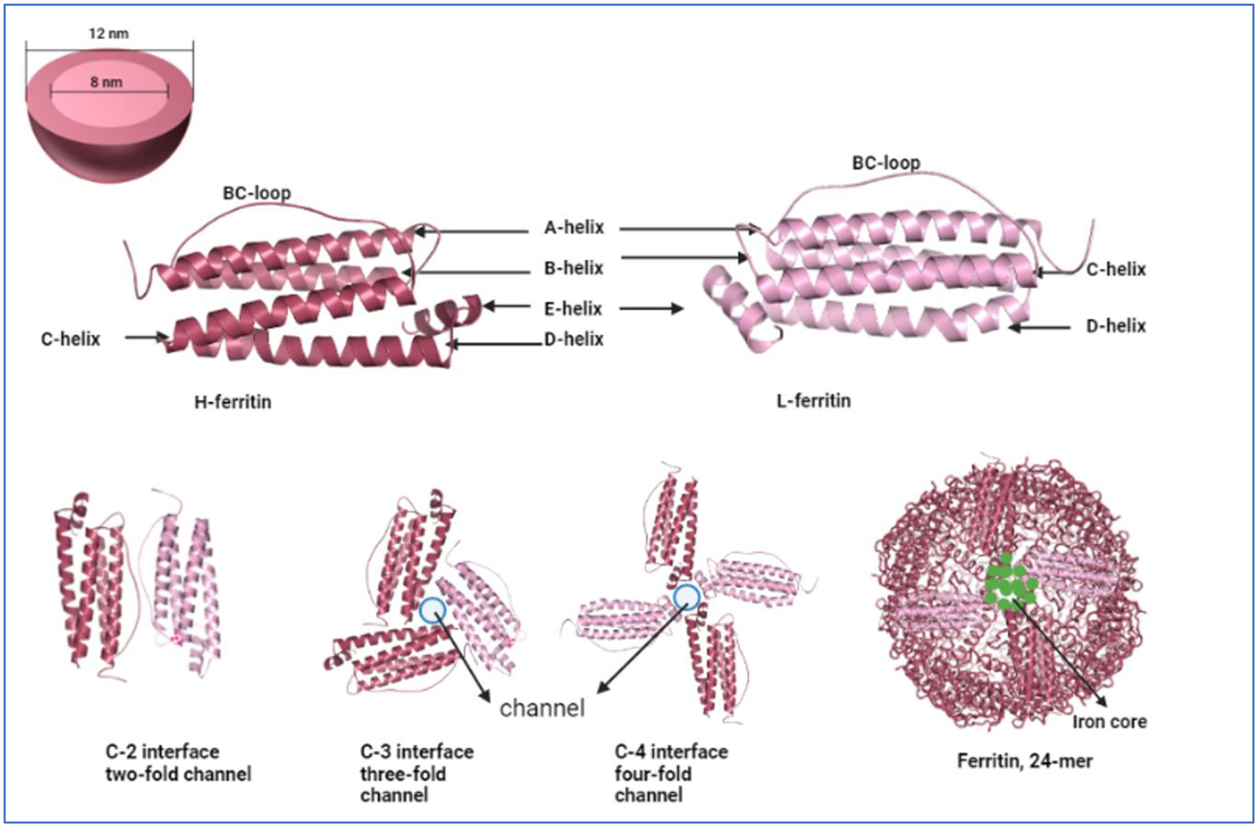
Fig. 1. Structure of Ferritin.

(A) A representation of ferritin as a sphere showing the inner core and outer core diameter. (B) Graphical representation of the identical ferritin H- and L- peptides. Images were built in PyMOL 2.5.4 using ferritin heavy chain PDB ID 1FHA and ferritin light chain PDB ID 2FFX. (C) The various interfaces of the heavy and light chain ferritin. The three- and four-fold channels represent the open channels through which iron atoms can flow in and out of the structure. Finally, a 24-mer representation of the ferritin structure containing iron atoms is also presented. The image is inspired from the review by Chenxi Zhang et al13.
