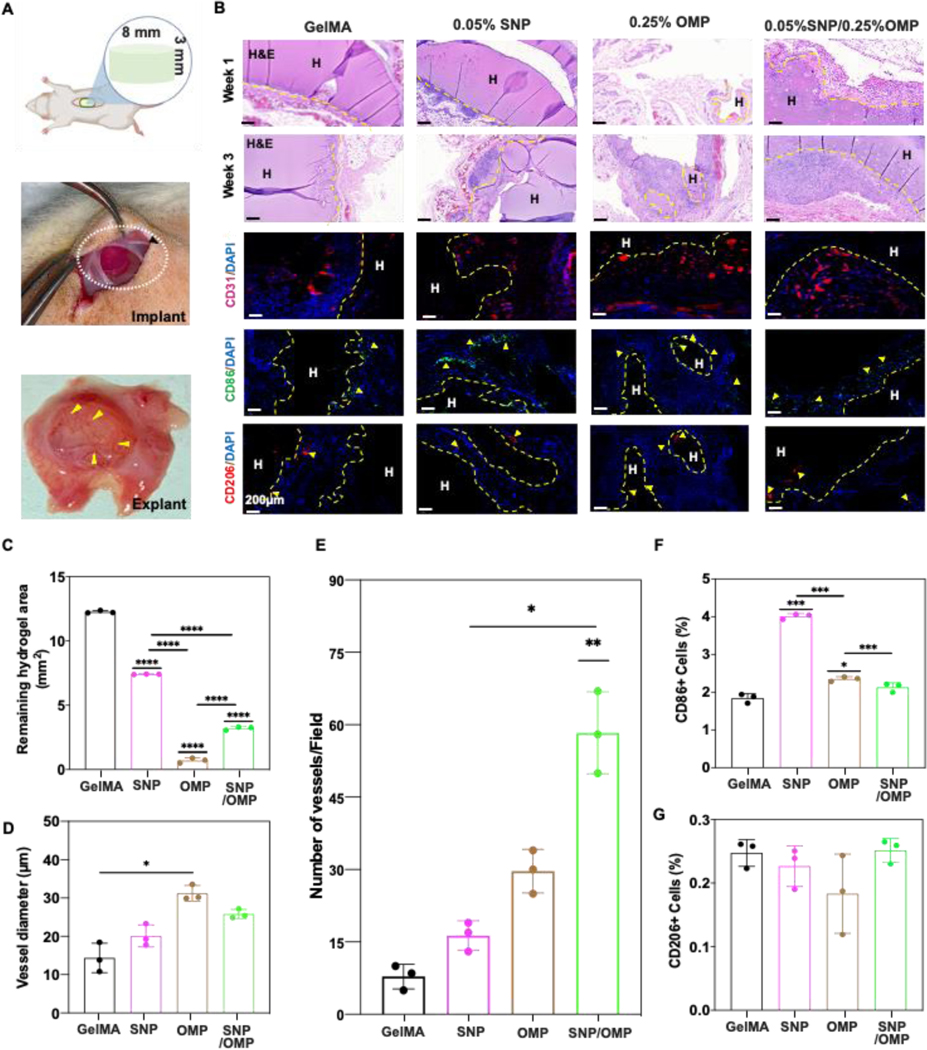
Figure 5: Biodegradation and biocompatibility of SNP/OMP hydrogels after subcutaneous implantation.
(A) Schematic representation and photograph depicting the procedure of subcutaneous implantation. (B) Microphotographs of histological sections stained with H&E staining 1- and 3 weeks post-implantation or immunohistochemically stained for neovascularization (CD31) or immune response (CD86 and CD206) of different hydrogels without cells 1- or 3-weeks post implantation. Groups: (1) GelMA, (2) SNPs with 0.05% SNP, (3) OMPs with 0.25% OMP, and (4), SNP/OMPs with 0.05% SNP+0.25%OMP. Semi-quantitative image analyses of (C) biodegradation of hydrogels (n=3; ****p<0.0001), (D) vessel diameter (n=3; ****p<0.0001), (E) number of vessels per field of view (n=3; ***p<0.001, **p<0.01, *p<0.05), (F) percentage of CD86+ cells, and (G) percentage of CD206+ cells three weeks post-implantation. (n=3; ***p<0.001, *p<0.05).

