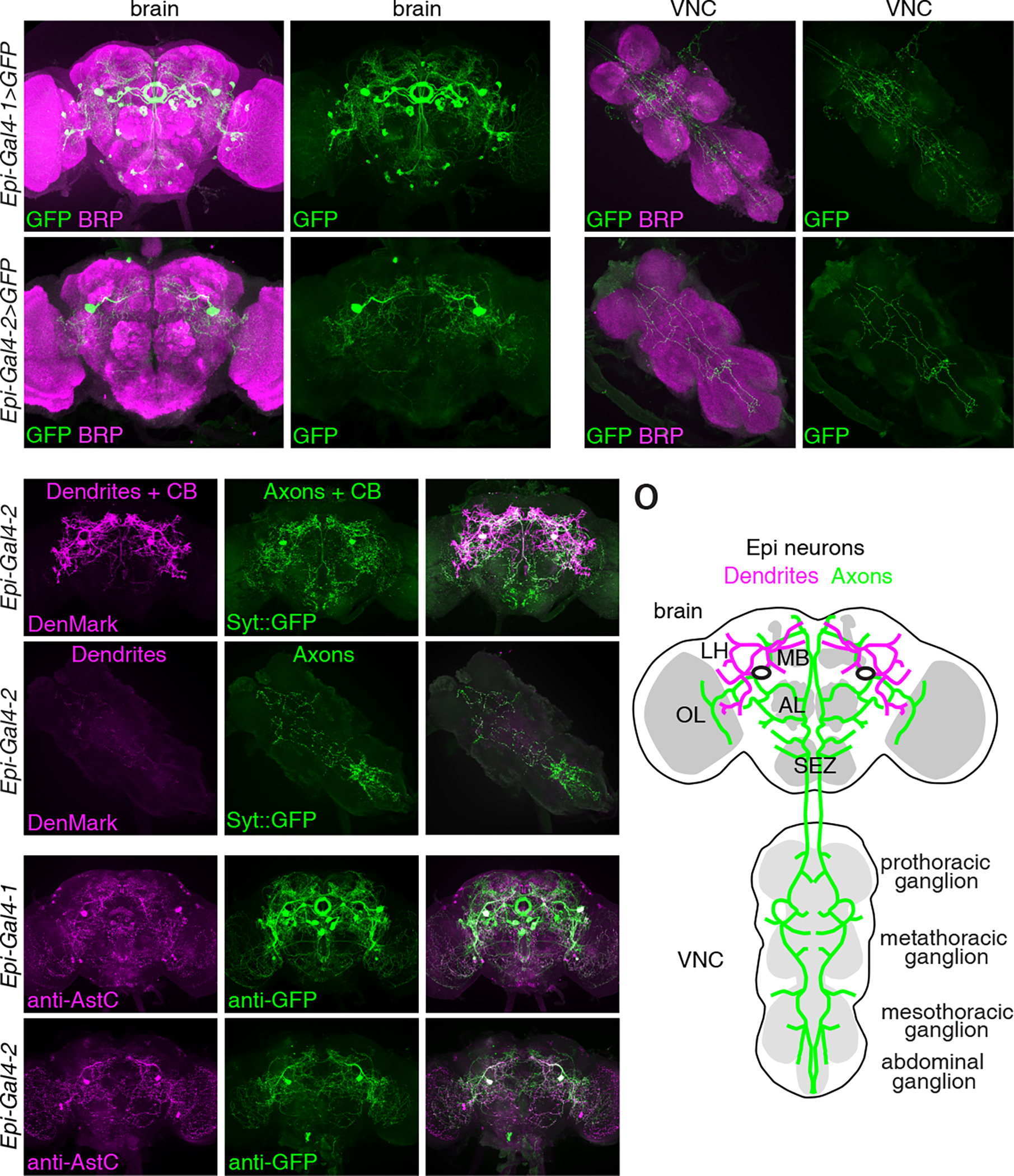
Figure 2. Spatial localization of Epi neurons.

(A-H) Expression patterns of Epi-Gal4-1 (A-D) and Epi-Gal4-2 (E-H) in the brain and VNC. Gal4-driven expression of UAS-mCD8::GFP is indicated in green (anti-GFP) and the neuropil marker BRP is labeled in magenta (anti-BRP). Scale bars indicate 50 μm. (I-N) Axons (green) and dendrites (magenta) of Epi-Gal4-2 positive neurons labeled with Syt::eGFP and DenMark, respectively. (I-K) Brain. (L-N) VNC. Scale bars indicate 50 μm. CB, cell body. OL, optic lobe. LH, lateral horn. MB, mushroom body. AL, antenna lobe. SEZ, subesophageal zone.
(O) Schematic illustration of the dendrites (magenta), axons (green) and cell bodies (two black ovals) of Epi neurons in the brain and VNC.
(P-U) Adult brains stained with anti-AstC (magenta) and anti-GFP (green). (P-R) Epi-Gal4-1-driven expression of UAS-mCD8::GFP. (S-U) Epi-Gal4-2-driven expression of UAS-mCD8::GFP. Scale bars indicate 50 μm.
See also Figure S3.
