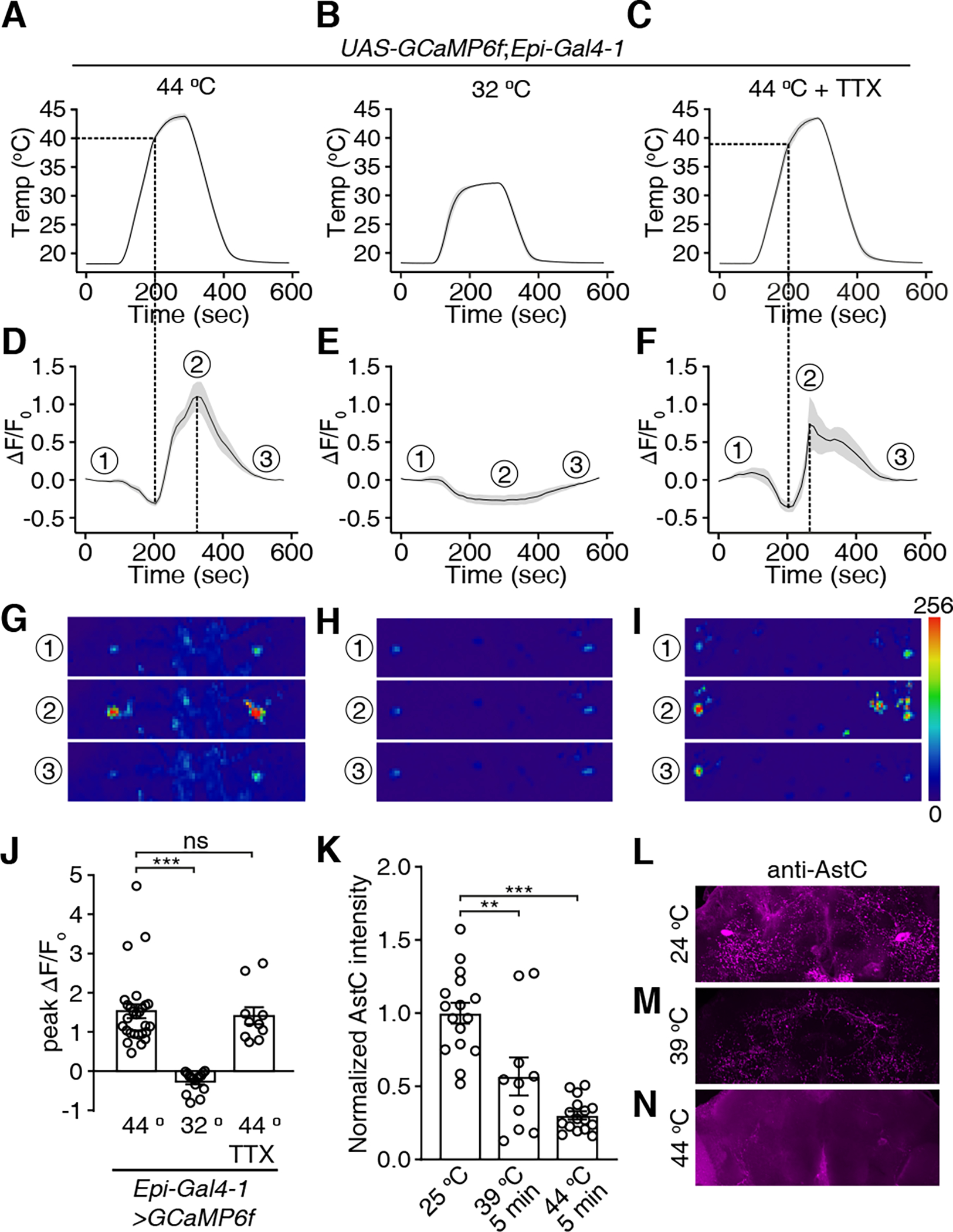
Figure 4. Epi neurons are direct sensors for nociception.

Ca2+ signals of Epi neurons during temperature changes. UAS-GCaMP6f was expressed under control of the Epi-Gal4-1. Brains were dissected and GCaMP6f signals were monitored in response to temperature ramps in the absence or presence of tetrodotoxin (TTX).
(A-C) Temperature ramps with the maximum temperatures indicated. TTX was added to the bath in (C).
(D) Changes in GCaMP6f signals (ΔF/F0) in response to the temperature ramp in (A). The ①, ②, and ③ indicate the time points for the sample images in (G-I).
(E) Changes in GCaMP6f signals (ΔF/F0) in response to the temperature ramp in (B).
(F) Changes in GCaMP6f signals (ΔF/F0) in response to the temperature ramp in (C) in the presence of TTX.
(G-I) Images of the GCaMP6f signals in Epi neurons (the dashed circles indicate the cell bodies) at three time points during the Ca2+ imaging as indicated in (D-F), respectively. Scale bars indicate 50 μm.
(J) Average peak ΔF/F0 exhibited by Epi neurons in (A-C). n = 10–27 neurons from ≥6 dissected brains. Error bars indicate S.E.M.s. Mann-Whitney test. ***P < 0.001, ns, not significant.
(K) Normalized anti-AstC signals in Epi neurons. The brains were incubated at room temperature (~ 24 °C), at 39 °C, or at 44 °C for 5 minutes. n = 10–16 neurons from ≥5 dissected brains. Error bars indicate S.E.M.s. Mann-Whitney test. **P < 0.01. (L-N) Sample images of anti-AstC signals at 25 °C (L), 39 °C (M) and 44 °C (N).
See also Figure S5.
