
Figure 5:
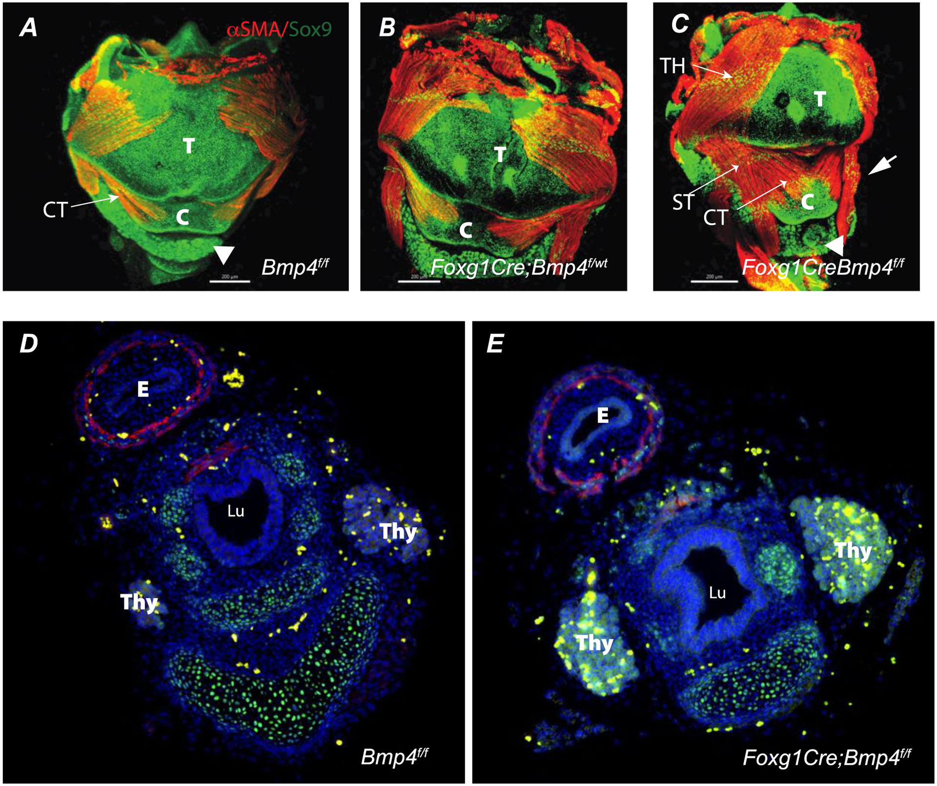
Whole mount immunofluorescence of larynx at E16.5 (A-C) are shown. A-C: Ventral view of control (A) heterozygous (B), and mutant (C) larynx. Cartilage and muscle were detected in all genotypes analyzed as determined by positive immunofluorescence detection for Sox9 (green) and αSMA (red), respectively. Note in the mutant at E16.5 the anomalous appearance of the muscle and cartilaginous elements of the larynx (arrow in C) and the lower, more distal position of the thyroid (arrowhead in C vs A control). Panels D and E depict cross sections of E16.5 esophagus and distal larynx/upper trachea. Note the anomalous shape of the airway in the mutant. . TH= thyrohyoideus muscle, ST= sternohyoideus muscle, CT= cricothyroid muscle, E= esophagus, T= thyroid cartilage, C = cricoid cartilage, . Thy= Thyroid, Lu= Lumen. Scale bar = 200 μm.