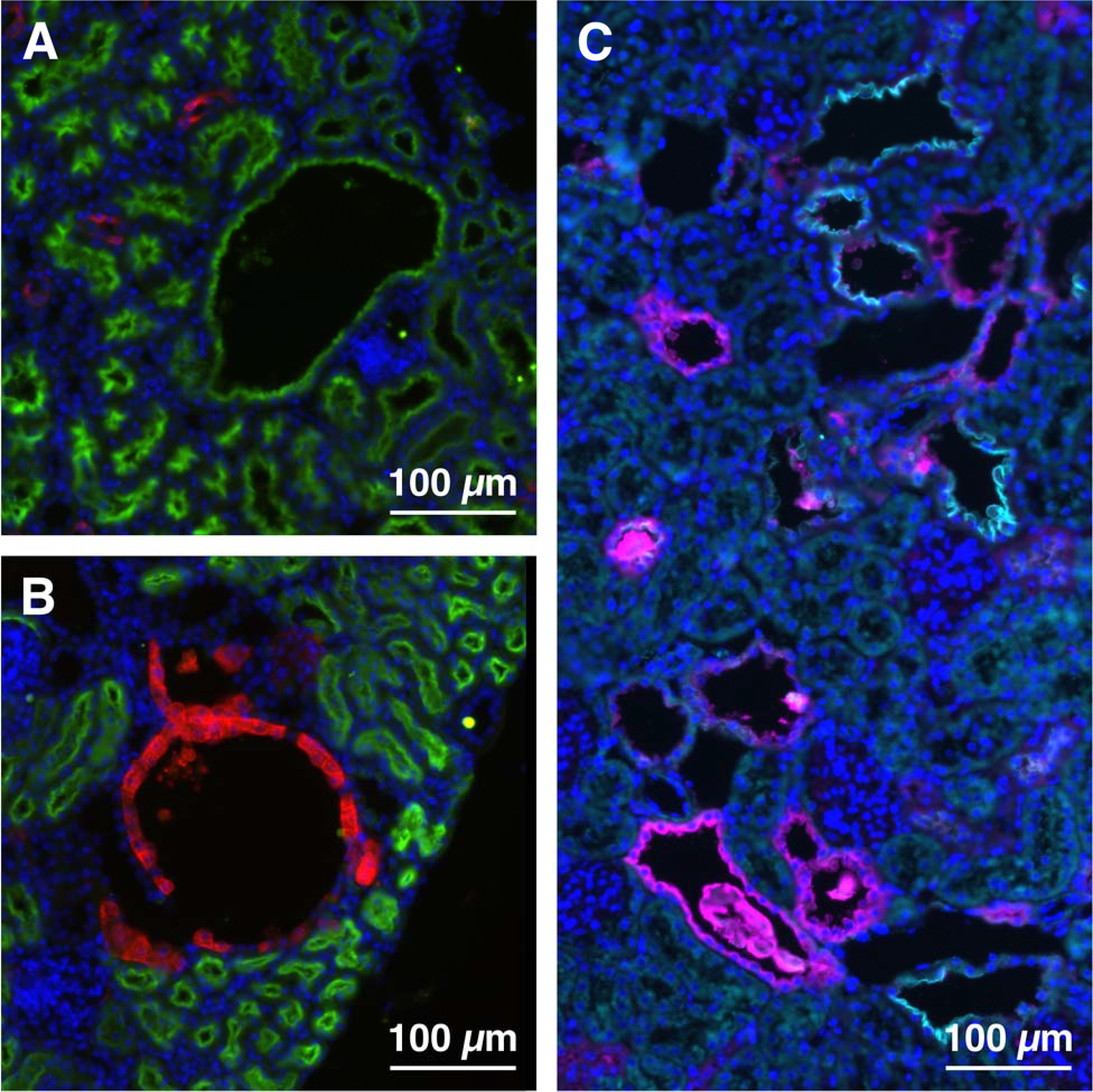
Figure 3. Cysts in Arl13bV358A/V358A kidneys are present in all nephron segments.

(A) LTL (proximal tubule, green), (B) DBA (collecting duct, red), (C) THP (thick ascending limb of the loop of Henle, magenta) and NCC (distal convoluted tubule, cyan) staining in Arl13bV358A/V358A kidney sections. Scale bars: 100 μm.
