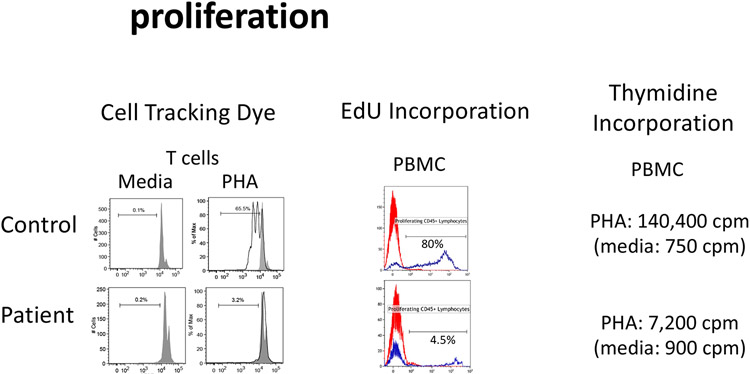
Figure 7: Cell proliferation.
The ability of lymphocytes to undergo proliferation can be analyzed in vitro. Cells are labelled with cell tracking dyes such as CFSE or cell trace violet which are evenly distributed to daughter cells following each round of division (evidenced by a decrease in fluorescence). Alternatively, cells are activated in vitro followed by evaluating the uptake of tritiated thymidine or analogue of tritiated thymidine, ethynyl deoxyuridine (EdU).

