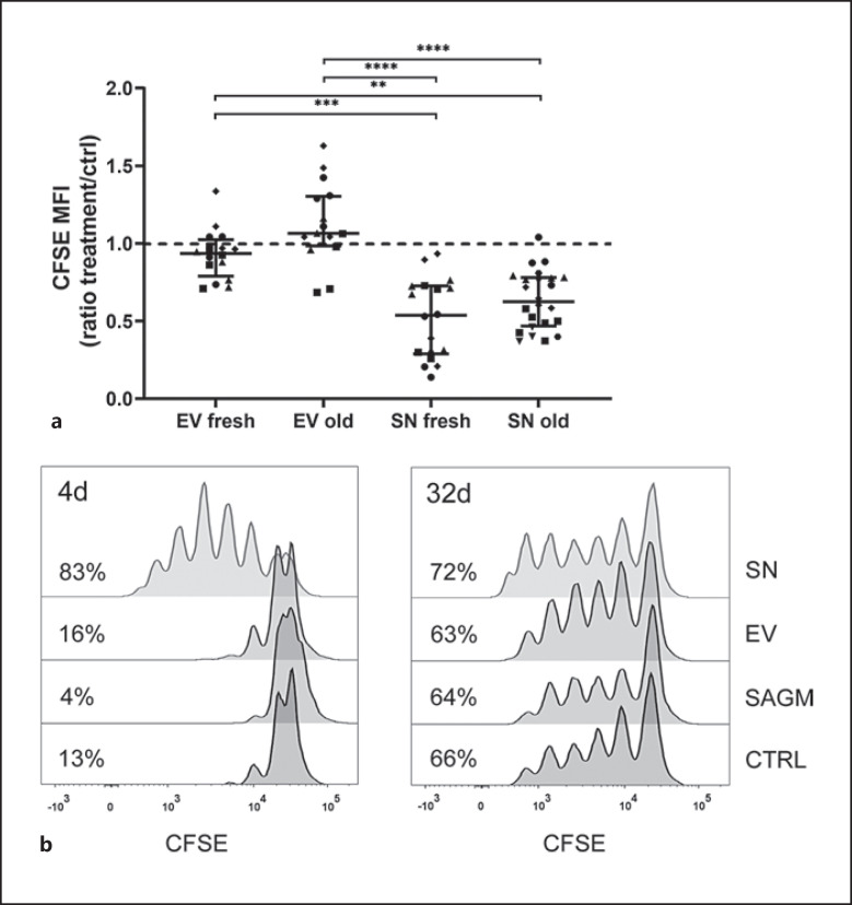
Fig. 2.
PBMCs were labeled with CFSE, stimulated with anti-CD3 and cultured for 4 days. CFSE is halved with each cell division; a lower CFSE MFI indicates augmented proliferation. a The data are presented as median (IQR). PBMCs exposed to fresh (n = 18) and longer stored RBC SN (n = 24) proliferated significantly more in relation to control (n = 20), while fresh (n = 16) and longer stored RBC EVs (n = 16) had varying effects on proliferation with no statistical significance in relation to control. Both fresh and old SNs increased cell proliferation significantly compared with fresh and old EVs and with control (Kruskal-Wallis test, p = 0.005 EV fresh vs. SN fresh, p = 0.007 EV fresh vs. SN old, p < 0.001 EV old vs. SN fresh and p < 0.001 EV old vs. SN old). RBC storage age was not significant. The number of samples differs among groups due to the data were extracted from several individual experiments. Symbols represent individual PBMCs. b Representative figure of T-cell proliferation of PBMCs exposed to SAGM, and SN and EVs from an RBC unit at days 4 and 32 of storage. Percentages of proliferating cells from lymphocyte population are shown. Individual PBMCs responded to anti-CD3 stimulation very differently and different level of proliferation was observed among experiments, which is seen. Thus, results are always compared to their own control. MFI, median fluorescence intensity.

