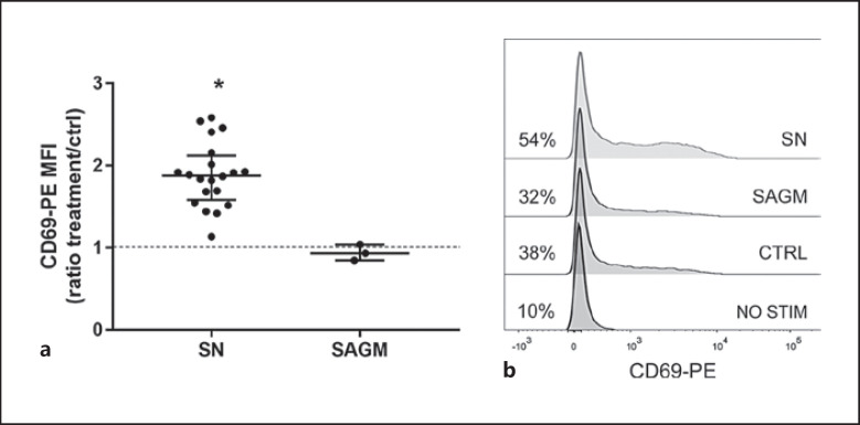
Fig. 3.
a Five-hour incubation with SN resulted in significantly increased expression of early activation marker CD69 in anti-CD3 activated T-cells (CD69-PE MFI values were compared with the Friedman test, p = 0.01 SN vs. ctrl). The data are shown as ratios to control and presented as median (IQR). b Representative examples of CD69 expression in non-activated, anti-CD3-activated, and SAGM- and SN-exposed anti-CD3-activated T cells. Percentages of positive cells from lymphocyte population are shown. CD69 gates were assigned based on isotype control.

