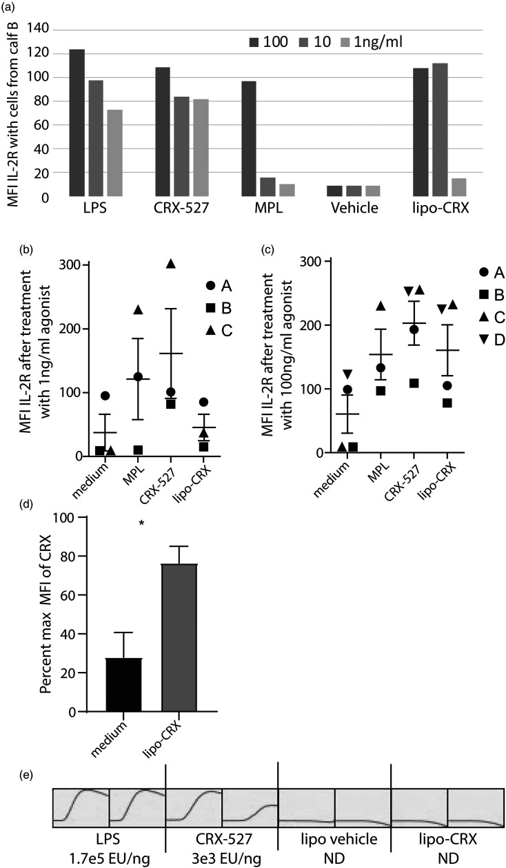
Figure 1.
Lipo-CRX activates bovine γδ T cells in mixed cultures in vitro, despite lack of LAL reactivity. a. Cells were treated with agonists as shown at varying concentrations. After 24 h, IL-2R expression was measured by flow cytometry on bovine γδ T cells. This assay was performed in single wells precluding statistical analyses. This procedure was repeated on cells from 3 (1 ng/ml) or 4 (100 ng/ml) different bovine calves at two concentrations of agonist. b. 1 ng/ml and c. 100 ng/ml. Standard error bars are shown. d. To normalize the data between experiments shown in Figure 1C, we calculated % of maximum, with the CRX value being the maximum value, for control and lipo-CRX treatment for each calf experiment. Data were pooled, means and SEM calculated and significance tested by unpaired Student's t-test. e. LPS, CRX-527, lipo-CRX and vehicle only control (liposomes without CRX-527) were compared in an LAL assay in duplicate showing development of Endotoxin Units (EU) over time and EU values as calculated by comparison to a standard curve are shown for each. LPS was strongly reactive, CRX-527 less so, and lipo-CRX had no reactivity and appeared similar to vehicle only (ND-not detected).

