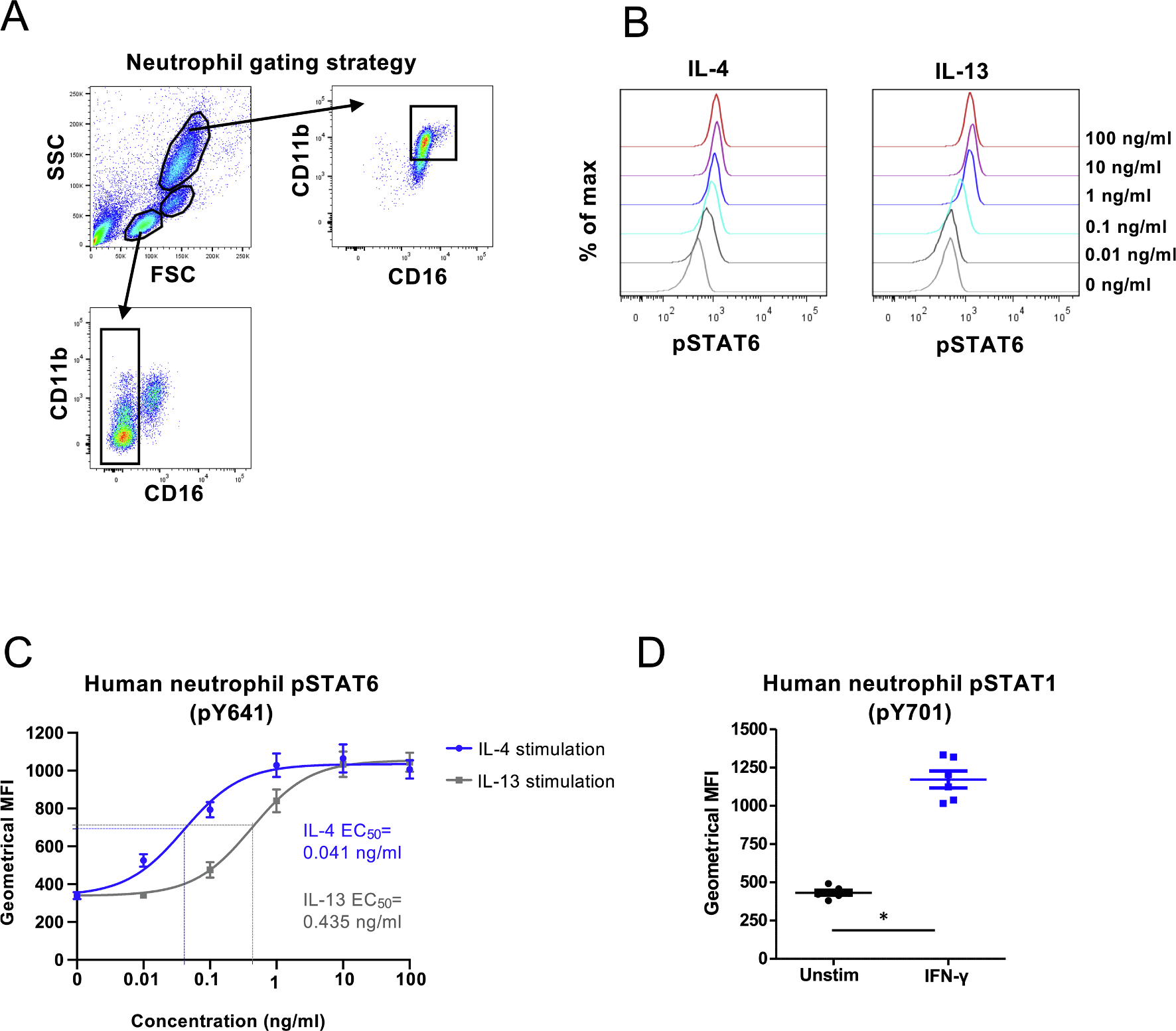
Fig. 1. Responsiveness of human neutrophils to 15 min stimulation with IL-4, IL-13 and IFN-γ, measured by phosphorylation of STAT6 and STAT1 using flow cytometry.

(A) Gating strategy for neutrophils, monocytes and lymphocytes. Neutrophils were defined as CD11bhigh CD16high cells. Monocytes and lymphocytes were gated based on forward and side scatter characteristics and CD16 + cells were excluded from cells in lymphocyte gate. Doublets were excluded from all three populations (not shown). (B) Stimulation of human whole blood was performed with indicated amounts of IL-4 or IL-13 and phosphorylation of STAT6 was determined in CD11bhighCD16high cells. Representative histograms of fluorescence intensity are shown. (C) Geometrical means ± SEM of pSTAT6 fluorescence of six donors after stimulation with IL-4 or IL-13. The curves of both stimulations are fitted using non-linear regression 3-parameter fit (goodness of fit was evaluated with R square and the values were for IL-4 and IL-13 0.8604 and 0.8961, respectively) and EC50 values are indicated based on the fitted curves. The IL-4 10 ng/ml datapoint was lost for one donor. (D) Geometrical means of pSTAT1 (pY701) fluorescence of six donors after whole blood was either left untreated or stimulated with 100 ng/ml of IFN-γ for 15 min, *) p = 0,0313; Wilcoxon matched-pairs signed rank test.
