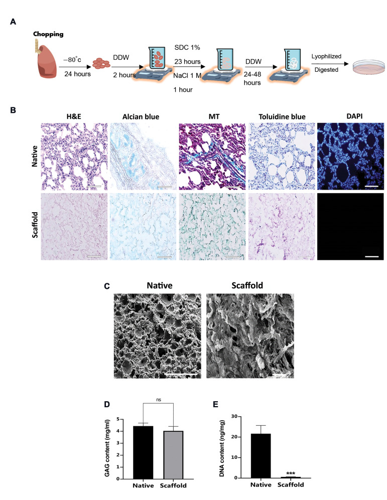
Fig.1.
Decellularization procedure, hydrogel formation and characterization. A. Schematic representation of lung decellularization procedure and hydrogel formation, B. Histological analysis of native sheep lung and decellularized sheep lung ECM-derived scaffold using H&E, Alcian blue, MT, Toluidine blue and DAPI staining (n=3 biological repeats, scale bar: 50 µm), C. Representative SEM photomicrographs of native lung (scale bar: 200 µm) and decellularized lung ECM-derived scaffold (scale bar: 50 µm). D. Quantification of GAGs content of native and decellularized scaffold (n=4). E. DNA quantification assay (n=3). DDW; Double distilled water, SDC; Sodium deoxycholate, PBS; Phosphate buffered saline, MT; Masson’s trichrome, SEM; scanning electron microscopy, ECM; Extracellular matrix, GAGs; glycosaminoglycans, h; Hour, ns; Not significant, and ***; P<0.001.

