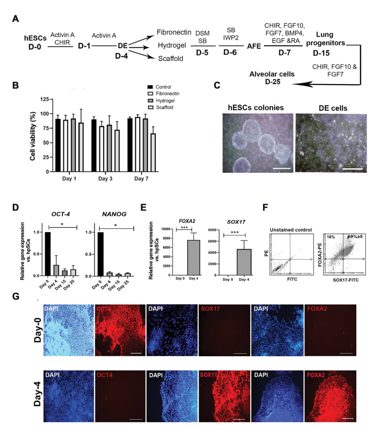
Fig.2.
Characterization of hESC-derived definitive endoderm cells at day 4 of differentiation. A. Schematic representation of lung differentiation protocol showing differentiation of hESCs into DE cells in the presence of small molecules and growth factors and replating lung progenitors on the different beds including fibronectin, hydrogel, and dECM at day 15. B. Determining the cell viability percentage by the MTS assay at days 1, 3, and 7 after the A549 cell line culture on the different beds. A549 cells were cultured as a control group (n=4). C. Phase-contrast images of typical hESC colonies (scale bar: 500 µm) and DE cells (scale bar: 200 µm) before replating. D. qRT-PCR analysis of hPSCs pluripotent marker genes at days 4, 15, and 25 of lung progenitor differentiation. E-G. Characterization of hPSC-derived DE cells before replating on fibronectin: E. qRT-PCR analysis for FOXA2 and SOX17 genes at day 4 of differentiation. F. Flow cytometrically analysis of FOXA2 and SOX17 positive cells (n=3). G. Evaluation of OCT4, FOXA2, and SOX17 protein expression in both hESC (upper panel scale bar: 500 μm, 200 μm, 500 μm, respectively) and DE cells (lower panel: 500 μm) by immunostaining. hESC; Human embryonic stem cell, dECM; Decellularized extracellular matrix, D; Day; DE; Definitive endoderm, AFE; Anterior foregut endoderm, hPSC; Human pluripotent stem cell, qRT-PCR; Quantitative real-time-polymerase chain reaction, *; P<0.005, and ***; P<0.001.

