
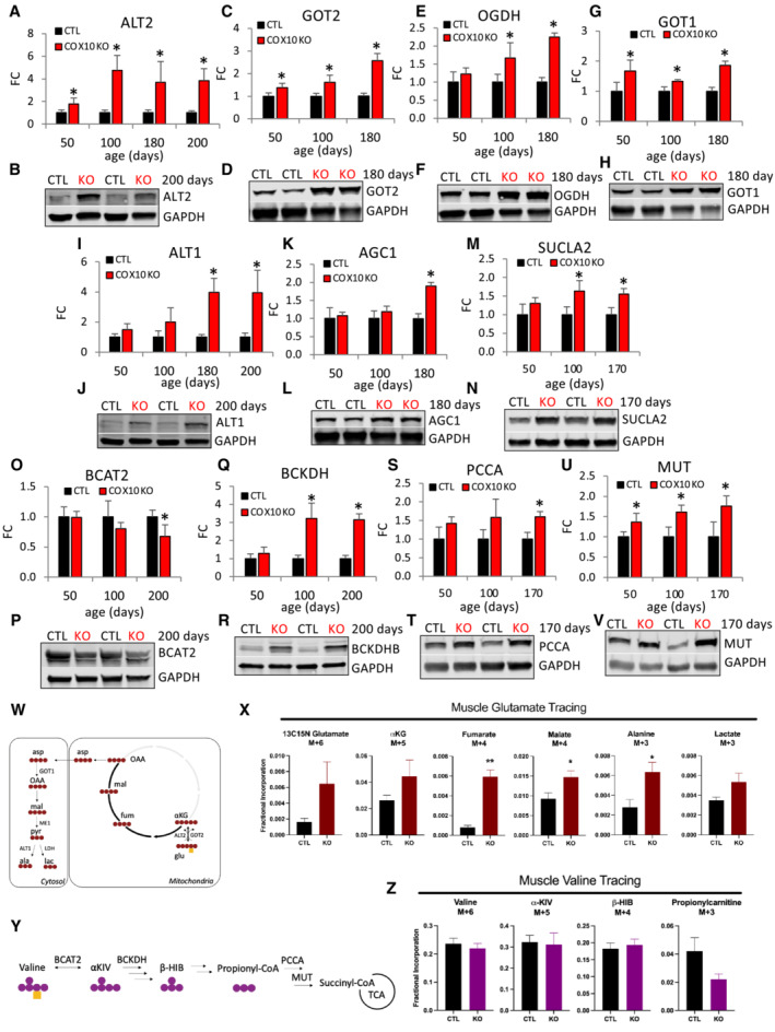
Figure 2. Upregulation of key components of amino acid oxidation progresses with age in COX10 KO muscle.
-
A–VAge‐dependent muscle protein levels of ALT2 (A), GOT2 (C), OGDH (E), GOT1 (G), ALT1 (I), AGC1 (K), SUCLA2 (M), BCAT2 (O), BCKDH (Q), PCCA (S), and MUT (U), estimated by band densitometry normalized by GAPDH, in COX10 KO expressed relative to same age CTL set at 1. Representative western blots of muscle lysates from 170‐ to 200‐day‐old mice separated by denaturing SDS–PAGE and probed for ALT2 (B), GOT2 (D), OGDH (F), GOT1 (H), ALT1 (J), AGC1 (L), SUCLA2 (N), BCAT2 (P), BCKDHB (R), PCCA (T), and MUT (V) and GAPDH (B, D, F, H, J, L, N, P, R, T, V).
-
WSchematic representation of oxidative glutamate‐αKG flux. Red circles represent 13C atoms and yellow square represents 15N atom derived from [13C5, 15N]‐glutamate. αKG, α‐ketoglutarate; OAA, oxaloacetate.
-
XMuscle fractional incorporation of M + 6 of glutamate, M + 5 of αKG, M + 4 of fumarate, M + 4 of malate, M + 3 of alanine, and M + 3 of lactate, in 200 days COX10 KO and CTL mice IP injected with [13C5, 15N]‐glutamate.
-
YSchematic representation of valine oxidative pathway. Purple circles represent 13C atoms and yellow square represent 15N atom derived from [13C5, 15N]‐valine. α‐KIV, α‐ketoisovalerate; β‐HIB, β‐hydroxyisobutyrate.
-
ZMuscle fractional incorporation of M + 6 of valine, M + 5 of α‐KIV, M + 4 of β‐ΗΙΒ, M + 3 of propionyl carnitine, in 200 days COX10 KO and CTL mice IP injected with [13C5, 15N]‐valine.
Data information: In panels (A, C, E, G, I, K, M, O, Q, S, and U), data are presented as Mean ± SD. COX10 KO (n = 4), CTL (n = 4). In panels (X and Z), data are presented as Mean ± SEM. In panel (X), COX10 KO (n = 4), CTL (n = 4). In panel (Z), COX10 KO (n = 3), CTL (n = 3). Statistically significant differences between the two groups were estimated by unpaired two‐tailed Student's test. *P < 0.05, **P < 0.005 COX10 KO versus CTL.
Source data are available online for this figure.