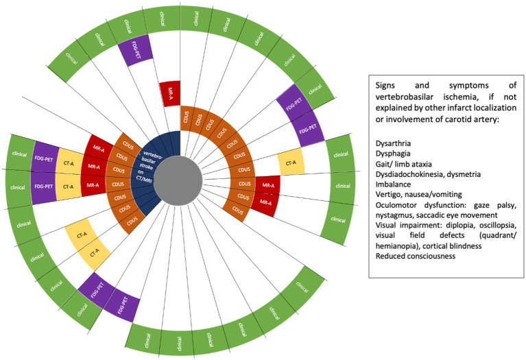
Figure 2.
Patients with giant cell arteritis (GCA) and vertebral artery (VA) involvement (n = 29). FDG-PET, 18F-fluorodeoxyglucose positron emission tomography; CT-A, computed tomography angiography; MR-A, magnetic resonance angiography; CDUS, colour Doppler ultrasound. Each ray reflects one patient.

