. 2023 Jul 10;13(30):20495–20511. doi: 10.1039/d3ra02248j

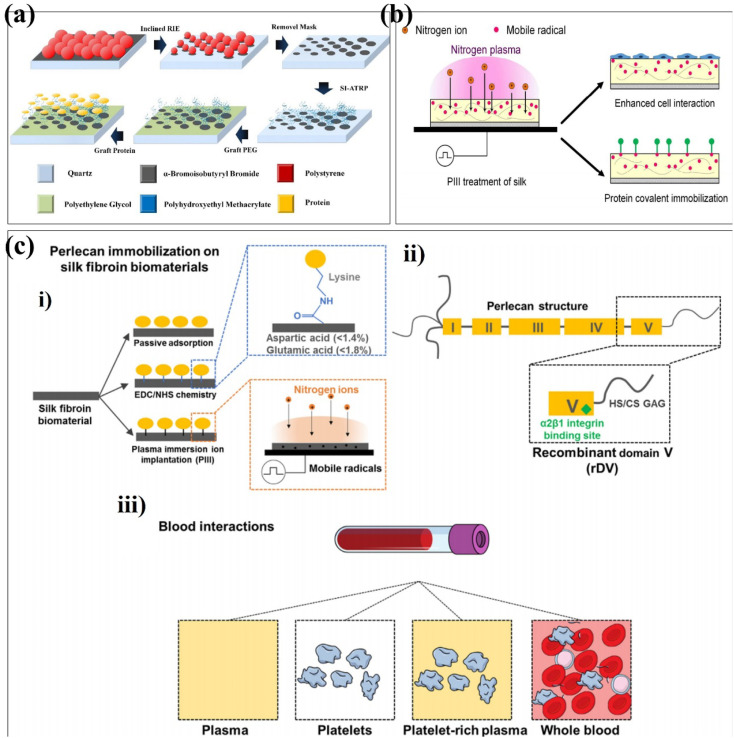
Fig. 7. Schematic depiction of plasma strategies onto surface-formed multifunctional biomaterials. (a) Schematic illustration of the fabricating process of graded protein/PEG nanopattern arrays. (b) Schematic of the fabrication process of surface modification using plasma immersion ion implantation (PIII). (c) Schematic of silk biofunctionalization with recombinantly expressed domain V (rDV) of human perlecan for blood contacting applications. (i) Schematic of the silk film biofunctionalization with rDV via passive adsorption (silk), carbodiimide chemistry (EDC/NHS silk) or nitrogen plasma immersion ion implantation (PIII silk). EDC/NHS-based protein immobilization occurs via formation of an amide bond between a carboxylic acid and a primary amine, typically occurring between aspartic/glutamic acid on silk and lysine on rDV. PIII-based immobilization relies on diffusion of mobile radicals from the PIII-treated silk surface to react with rDV. (ii) Schematic of the main structural and functional features of perlecan domain V expressed recombinantly in HEK-293 cells. rDV is a proteoglycan consisting of an 80 kDa protein core decorated with either a heparan sulfate (HS) or a chondroitin sulfate (CS) glycosaminoglycan (GAG) chain and has an α2β1 cell integrin binding site located at the C-terminus of the proteoglycan. (iii) Schematic of blood contacting assays used in this study with an increasing amount of blood components and complexity of interaction with the exposed surface. These are reprinted with permission from Xue et al. (2019, ACS Appl. Mater. Interfaces), Kondyurin et al. (2018, ACS Appl. Mater. Interfaces), Lau et al. (2021, Acta Biomater.), respectively.

Fig. 7