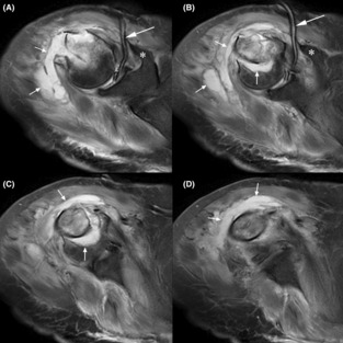
FIGURE 2.

Sequential T2 (fluid enhancing) MR images in coronal view (A is most proximal, D is most distal). Asterisks (*) indicate the coracoid process, longer arrows indicate a Jackson–Pratt drain within the glenohumeral joint (A, B), and shorter arrows indicate the abscess. The abscess clearly extends into the medullary region of the bone at the fracture site (e.g., vertical arrows in B and C). The scan was obtained 13 days after the fracture.
