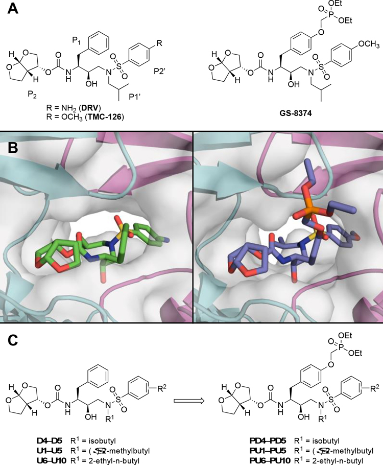
Figure 1.
Structures of HIV-1 protease inhibitors. (A) Darunavir (DRV), TMC-126, and the corresponding P1 phosphonate analog GS-8374. (B) DRV and GS-8374 bound to wild-type HIV-1 protease (PDB: 6DGX and 2I4W, respectively). The protease is depicted as a gray surface and a cartoon representation, with chain A in teal and chain B in magenta. (C) DRV analogs with variations at the P1′ and P2′ positions and the corresponding P1 phosphonate analogs analyzed in this study.

