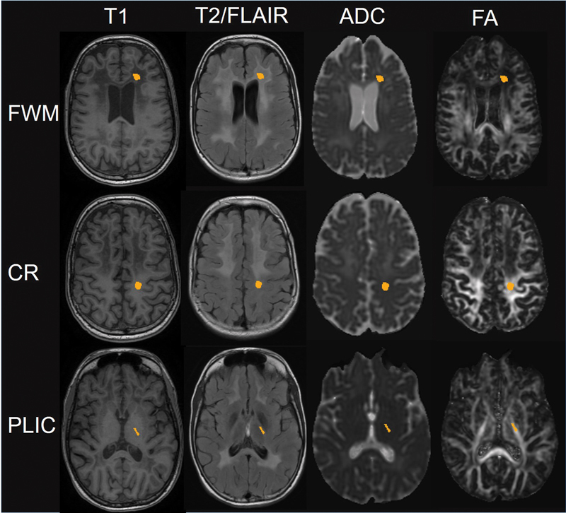
Fig. 1.

Exemplary illustration of the location of the regions of interest (ROIs) in the frontal white matter (FWM), the central region (CR), and the posterior limb of the internal capsule. Rows from left to right show the positioning of the ROIs in the anatomical images T1 and T2/FLAIR—these ROIs were transferred to co-registered DWI maps ADC and FA to derive the values within each ROI as mean of all marked voxels.
