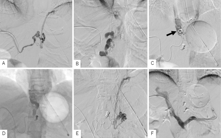
Figure 3.
Percutaneous transhepatic obliteration. A: Percutaneous transhepatic portal angiography revealed a branch of the right gastric vein. Coil embolization was performed from the microcatheter. B: A developed right gastric vein was visualized. It was considered to be a trunk of the varices continuing to the site of bleeding. C: Coil embolization was performed from the microcatheter advanced to the periphery of the right gastric vein to decrease the blood flow (arrow). D: EO (5%) was injected with the S-B tube blocking the drainage channel, and sufficient stagnation was obtained. E: A donor vein branching from the left gastric vein was also observed. Coil embolization was performed. F: Portal angiography after PTO on three donor channels confirmed their disappearance. EO: ethanolamine oleate, PTO: percutaneous transhepatic obliteration, S-B tube: Sengstaken-Blakemore tube

