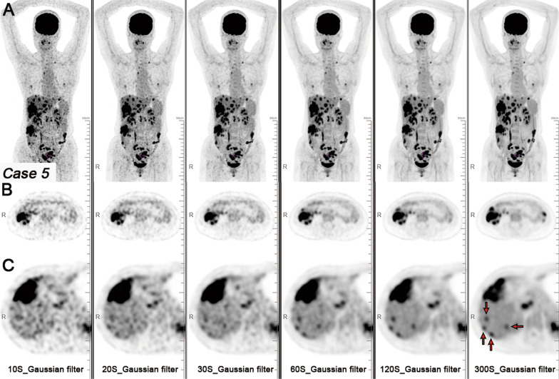
Fig. 4.
A patient with CRC and liver metastases. Maximum intensity projection (MIP) PET images (A), axial PET images of colon cancer (B), and axial PET images of liver metastases (C) with Gaussian filter with different acquisition durations (10, 20, 30, 60, 120, and 300 s). The 10-, 20-, and 30-s acquisition durations images with Gaussian filter exhibited noticeable noise, making it difficult to observe small liver metastases (red arrow). As the acquisition duration was extended, the liver metastases were clearly displayed

