Figure 2.
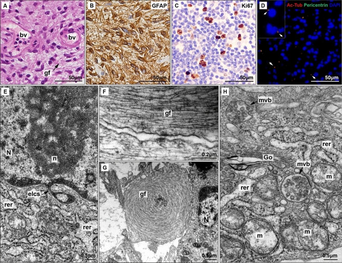
Histopathological and ultrastructural analysis of low-grade glioma. (A) HE staining shows increased cellularity with predominant gliofibrillar (gf) component. Blood vessels (bv) show non-hyperplasic endothelial cells. (B) GFAP and (C) Ki67 immunostaings. (D) Immunofluorescence; acetylated β tubulin (red) is a marker of axoneme while pericentrin (green) is present in basal body. Colocalisation of both markers show clearly the presence of primary cilia (arrows). (E) Electron micrograph of a nuclear envelope-limited chromatin sheets (ELCS) isolating heterochromatin (N: nucleus, n: nucleoli, rer: rugous endoplasmic reticulum). (F) Electron micrograph of abundant parallel gliofilaments (gf) along an astrocytic prolongation. (G) Electron micrograph of abnormal disposition of gliofilaments acquired a characteristic concentric shape. (H) Electron micrograph of multivesicular bodies (mvb) present in glioma astrocytes (Go: Golgi apparatus, m: mitochondria).

