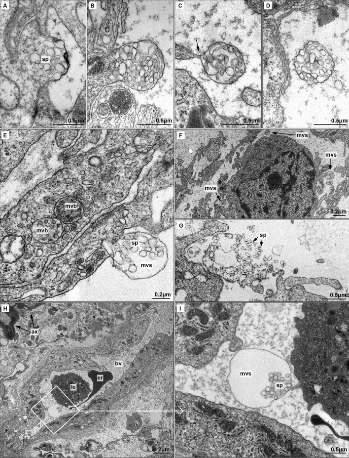
Figure 3.
Biogenesis and local and distant migration of MVSs (A–D) Biogenesis of MVS. (A) Spheresomes (sp) aggregate in a specific region of the cell membrane. Cell membrane begins to evaginate pushed by spheresomes. (B) A membrane evagination originates the multivesicular sphere (mvs), fulfilled by spheresomes. (C) The cell releases the MVS by narrowing the cell membrane (arrow). MVSs frequently appear along cytoplasmatic prolongations. (D) MVSs are released into the extracellular medium. (E–G) Short distance migration of MVSs and spheresomes. (E) Comparison of exosomes-loaded multivesicular bodies (mvb) and early MVS. Differences in the size of both multivesicular structure and inside vesicles are shown. (F) Panoramic view of MVSs surrounding a tumoural astrocyte. (G) Rupture of MVS membrane releases spheresomes (H–I). Long distance migration of MVSs and spheresomes. (H) Small blood vessel (bv) and nearby myelin-ensheathed axons (ax). An erythrocyte (er) and a leukocyte (le) are observed in the lumen of the vessel. (I) Detail from (H) showing an MVS with a dilated membrane in the lumen of a vessel. A small extension of the endothelial cell seems to guide the MVS.

