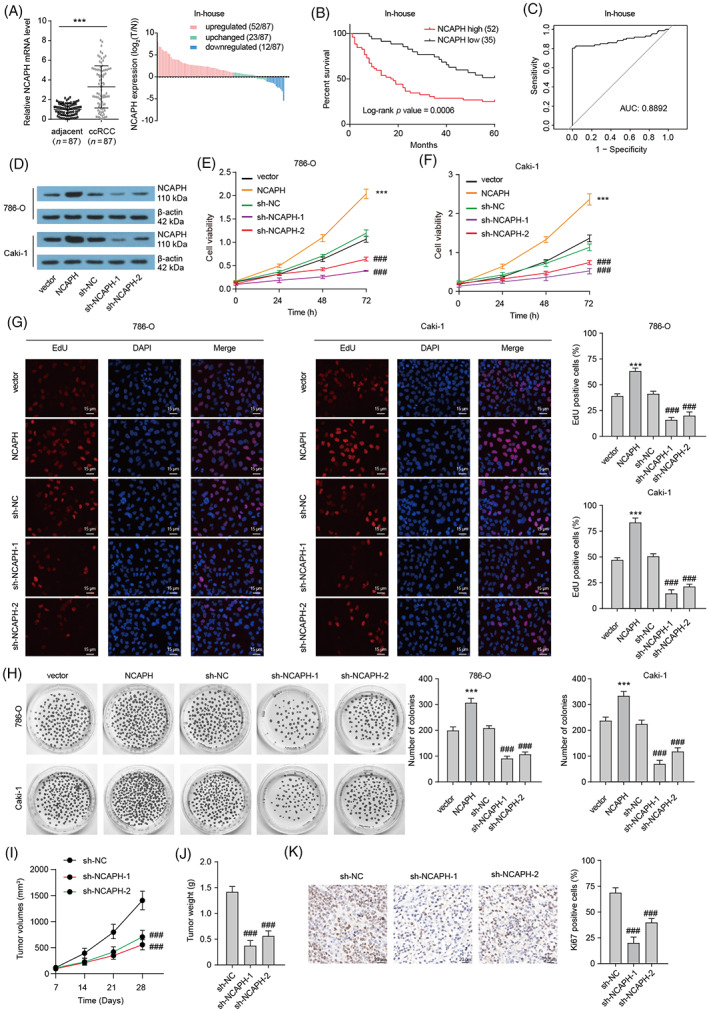
FIGURE 1.

Elevated non‐SMC condensin I complex subunit H (NCAPH) promotes clear cell renal cell carcinoma (ccRCC) growth. (A) NCAPH expression in 87 paired ccRCC tissues and normal tissues was assessed by qRT‐PCR. Paired t‐test been used, ***p < 0.001. (B) Kaplan–Meier plot showing the relationship between NCAPH levels and patient overall survival. (C) ROS curve showing the potential diagnostic value in ccRCC. (D) NCAPH protein expression in 786‐O and Caki‐1 cells with NCAPH overexpression or depletion was assessed by Western bolting. (E and F) Cell viability of 786‐O and Caki‐1 cells with NCAPH overexpression or depletion was assessed by CCK‐8 assays. ***p < 0.001 versus vector; ### p < 0.001 versus sh‐NC. (G) EDU staining shows the proliferative activity of 786‐O and Caki‐1 cells. ***p < 0.001 versus vector; ### p < 0.001 versus sh‐NC. (H) Clone formation assay showing the clonal formation ability of 786‐O and Caki‐1 cells. ***p < 0.001 versus vector; ### p < 0.001 versus sh‐NC. (I and J) Caki‐1 cells transfected with sh‐NC or sh‐NCAPH were injected into injected subcutaneously into Balb/c nude mice (five mice per group). The effect of NCAPH silencing on tumour volume curve and tumour weight was analysed. ### p < 0.001 versus sh‐NC. (K) Ki67 staining of xenografts. ### p < 0.001 versus sh‐NC. All experiments were repeated at least three times.
