Figure 2.

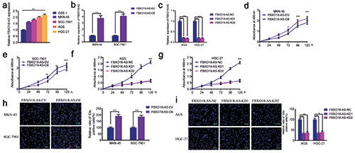
FBXO18-AS promoted gastric cancer cell proliferation. a) FBXO18-AS expression in different gastric cancer cell tissues was determined by qPCR (p<0.001; one-way ANOVA). b,c) qPCR validated FBXO18-AS expression in MKN-45 and SGC7901 cells after FBXO18-AS overexpression and in AGS and HGC-27 cells after FBXO18-AS knockdown (FBXO18-AS overexpression: p<0.001, Student’s t-test; FBXO18-AS knockdown: p<0.001; one-way ANOVA). d-g) MTS assays revealed that overexpression of FBXO18-AS impacted the viability of MKN-45 (d) and SGC7901 (e) cells, and FBXO18-AS knockdown affected the viability of AGS (f ) and HGC- 27 cells (g) (MKN-45: p<0.05; SGC7901: p<0.01; AGS: p<0.05; HGC-27: p<0.01; one-way ANOVA). h,i) EdU assays indicated that FBXO18-AS overexpression impacted the proliferation of MKN-45 and SGC7901 cells (h), and FBXO18-AS knockdown affected the proliferation of AGS and HGC-27 cells (i). Scale bars: 100 μm (MKN-45: p<0.001; SGC7901: p<0.001; Student’s t-test; AGS: p<0.001; HGC-27: p<0.001; one-way ANOVA). The overall data are presented as the mean ± SD (three independent tests);. ***p<0.001; **p<0.01; *p<0.05.
