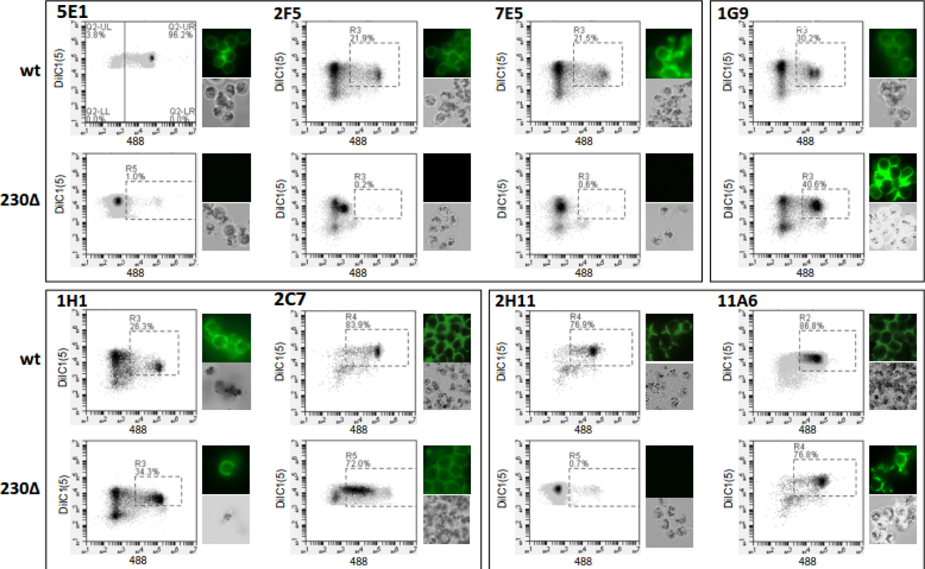
Figure 3.

Monoclonal (mAb) gamete/zygote surface reactivity. Purified wild-type strain NF54 (wt) or Pfs230Δ452–3135 (Pfs230Δ) gametes/zygotes were incubated with the indicated mAb, washed, and then stained with Alexa Fluor 488-labeled anti-mouse IgG and membrane potential dye DiIC1 (5). Fluorescence was monitored using flow cytometry and fluorescence microscopy. For each antibody and gamete/zygote population, the DiIC1(5) and Alexa Fluor 488 fluorescence of each cell is plotted and representative fluorescent and bright field images are shown.
