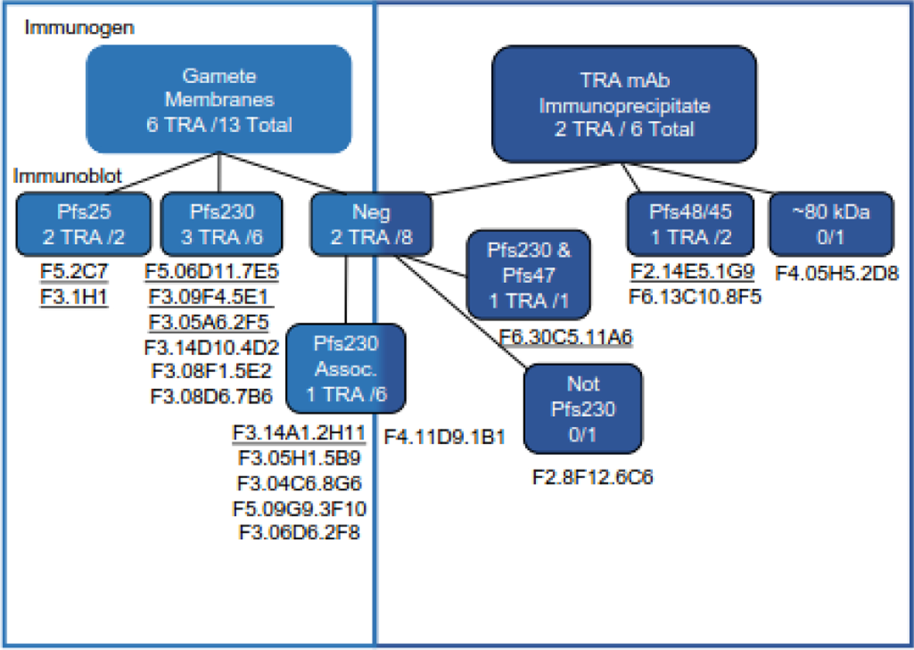
Figure 4.

Comparison of the profiles of monoclonals (mAbs) generated by each of the immunogens. mAbs that were derived from mice immunized with gamete/zygote membranes are in the light blue boxes and those from mice immunized with material immunoprecipitated by TRA mAbs are in the dark blue boxes. Within each box mAbs have been divided first based on their immunoblot reactivity and TRA mAbs are underlined. The immunoblot-negative mAbs are further separated based on their lack of reactivity with Pfs230Δ357–3135 gametes/zygotes (Pfs230 Assoc). The two immunoblot-negative mAbs that still reacted with Pfs230Δ357–3135 gametes/zygotes, 11A6 and 6C6, were then separated based on TRA and mass spectrometry data.
