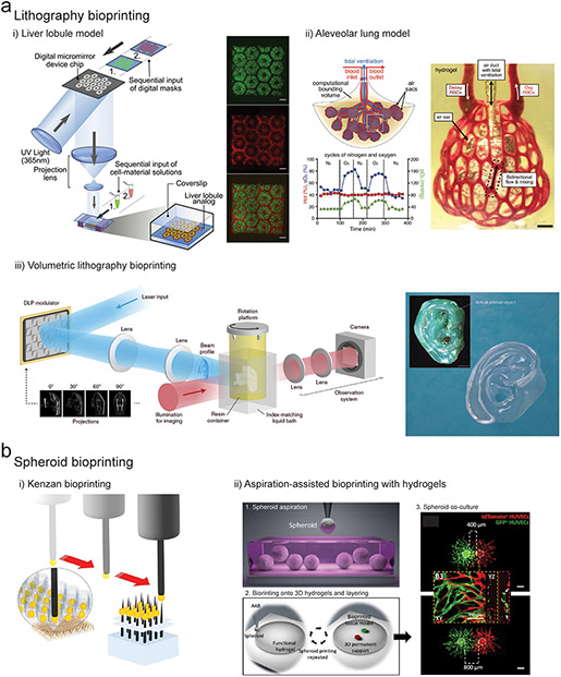
Figure 4. Advanced bioprinting technologies.
(a) i) DLP lithography bioprinting process where light is spatially projected onto a cell laden bioresin using a digital micromirror device to create a liver lobule construct (green regions contain iPSC-hepatocytes, and red regions contain endothelial cells & adipose-derived stem cells. Scalebars 500μm. (Ma et al., 2016) ii) DLP bioprinting of an alveolar lung model containing a central mechanically ventilated air sac surrounded by vascular channels perfused with red blood cells, and demonstration of gaseous exchange through measurement of reoxygenation of the red blood cell population (green line) following oxygenation of the air sac (blue line). Scalebars 1mm. (Grigoryan et al., 2019) iii) Experimental setup for volumetric bioprinting including laser input followed by DLP projection modulation of light onto a rotating platform containing the bioresin. Image of bioprinted human ear model created using a cell laden GelMA bioresin, total printing time 22.7 seconds. Scalebar 2mm. (Bernal et al., 2019; Loterie et al., 2020) (b) i) Kenzan bioprinting method where cell spheroids are aspirated and then skewered onto metal needles for fusion into 3D constructs. (Moldovan et al., 2016) ii) Aspiration-assisted bioprinting of spheroids (labelled red and green) onto fibrin hydrogels at different separation distances to study paracrine signaling and angiogenic sprouting. Scalebar 400μm. (Ayan et al., 2020)

