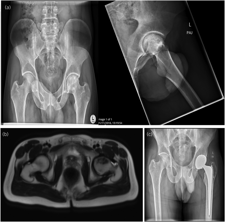
Figure 3 .
(a) Bilateral anteroposterior (AP) and left-sided lateral x-rays of the patient’s hip prior to hip decompression and stem cell injection showing the level of deterioration of the joint. (b) Bilateral MRI scan of the patient’s hips showing the progressive deterioration of the left hip. (c) Bilateral AP x-ray post left-sided total hip arthroplasty (THA).

