
Fig. 3.
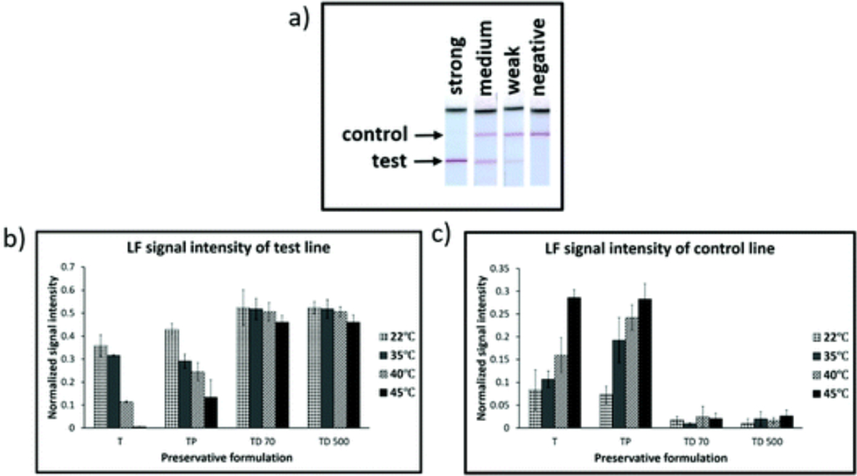
Performance of ldh1 iSDA of reagents stored dry in Std 17 GF for 360 h with different preservative formulations (Table 1), and range of temperatures. (a) Example of images of lateral flow (LF) detection strips showing strong, medium, weak, and negative amplification signal for ldh1 iSDA with 100 genomic copies. (b) and (c) Normalized intensities of LF test line and control line, respectively. Excellent stability was achieved for formulations that contained both trehalose and dextran (TD 70 and TD 500), and at all temperatures of storage as indicated by strong signal intensities for the test line and a weaker control line. With trehalose alone (T), target amplification failed with storage at 45 °C, and amplification was weaker for 22, 35, and 40 °C (when compared to TD 70 and TD 500) as indicated by weaker signal intensity for the test line and stronger intensity for the control lines. Samples with both trehalose and PEG (TP) performed only slightly better than trehalose alone.