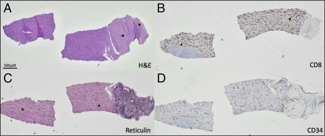
Figure 3.
Ectopic spleen. Hematoxylin and eosin-stained standard histologic sections show splenic tissue mainly composed of red pulp attached to a large hepatic portal tract (A). CD8 immunohistochemical stain labels littoral and endothelial cells with partial histiocytic functions specific to the spleen (B). Reticulin special stain highlights sinusoids (C). CD34 immunohistochemical stain shows normal blood vessels. Asterisks mark bile ducts. Arrowheads mark sinusoids.

