
Figure 1: Model of dynamic changes in ATMs in adipose tissue in response to overnutrition.
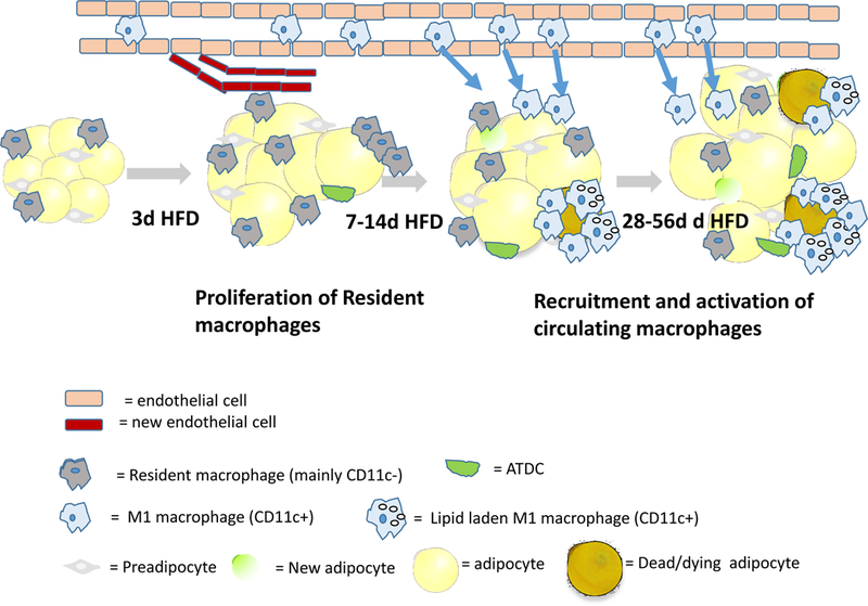
Muir et al found the HFD leads to a rapid (within 3d) expansion of adipose tissues of male mice and an increase in the proliferation of resident macrophages. Only later, as obesity develops, are circulating monocyte precursors recruited from the circulation into the tissue in a CCR2 dependent manner where they become activated (M1-like, CD11c+) and drive inflammation and tissue dysfunction. From 28–56 d of HFD, adipocyte size plateaus and CD11c+ ATMs accumulate lipid droplets. The magnitude and time course of these changes were more dramatic in eWAT than iWAT, but otherwise similar. The adipose expansion was due to increased adipocytes size, with only an early transient increase in preadipocyte proliferation and numbers, but this result does not preclude the possibility that some were differentiated into adipocytes, as illustrated. The results of Muir et al reinforce the importance of analyzing the dynamic changes in the cross-talk among leukocytes, endothelial cells and adipose progenitors in adipose tissue rsemodeling in response to acute and chronic overnutrition.